Journal of Fluorescence ( IF 2.6 ) Pub Date : 2021-09-21 , DOI: 10.1007/s10895-021-02814-0 T K Krishnapriya 1 , Ayswaria Deepti 2 , P S Baby Chakrapani 2, 3 , A S Asha 1, 3 , M K Jayaraj 4
|
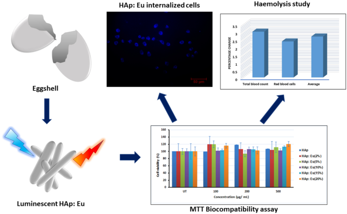
Hen’s eggshell, a biological waste product, was turned into a cell imaging probe: europium doped hydroxyapatite (HAp: Eu) nanoparticle using hydrothermal method. Luminescence of the synthesized nanoparticle was studied for various doping concentrations of the lanthanide ion europium (Eu3+). Eu doped HAp showed a hexagonal crystal structure and rod-shaped morphology. Well-defined emission peaks of europium, corresponding to the substitution of Eu3+ at the Ca2+(I) site of HAp, were confirmed from the samples’ photoluminescence (PL) spectra. Good biocompatibility up to 500 μg/mL of the samples indicates their potential applications in bioimaging. Synthesized nanoparticles were internalized and used for in vitro imaging of the PC12 cells without any surface modification. The materials’ use as a potential in vivo imaging agent is proposed from the haemolysis study.
Graphical Abstract
中文翻译:
用于细胞成像应用的蛋壳衍生铕掺杂羟基磷灰石纳米粒子
母鸡的蛋壳,一种生物废物,被转化为细胞成像探针:使用水热法掺杂铕的羟基磷灰石(HAp:Eu)纳米粒子。针对镧系离子铕(Eu 3+)的各种掺杂浓度,研究了合成纳米颗粒的发光。Eu掺杂的HAp显示出六方晶体结构和棒状形态。铕的明确发射峰,对应于Ca 2+处 Eu 3+的取代(I) HAp 的位点,由样品的光致发光 (PL) 光谱证实。高达 500 μg/mL 样品的良好生物相容性表明它们在生物成像中的潜在应用。合成的纳米粒子被内化并用于 PC12 细胞的体外成像,无需任何表面修饰。溶血研究提出了该材料作为潜在的体内显像剂的用途。


 京公网安备 11010802027423号