JACC: Cardiovascular Imaging ( IF 12.8 ) Pub Date : 2021-09-15 , DOI: 10.1016/j.jcmg.2021.07.023 Shuang Li 1 , Di Zhou 1 , Arlene Sirajuddin 2 , Jian He 1 , Jing Xu 1 , Baiyan Zhuang 1 , Jinghan Huang 3 , Gang Yin 4 , Xiaohan Fan 5 , Weichun Wu 6 , Xiaoxin Sun 7 , Shihua Zhao 1 , Andrew E Arai 2 , Minjie Lu 4
|
Objectives
The aim of this study is to examine the prognostic value of T1 mapping and the extracellular volume (ECV) fraction in patients with dilated cardiomyopathy (DCM).
Background
Patients with DCM with functional left ventricular remodeling have poorer prognoses. Noninvasive assessment of myocardial fibrosis using T1 mapping and the ECV fraction may improve risk stratification of patients with DCM; however, this has not yet been systematically evaluated.
Methods
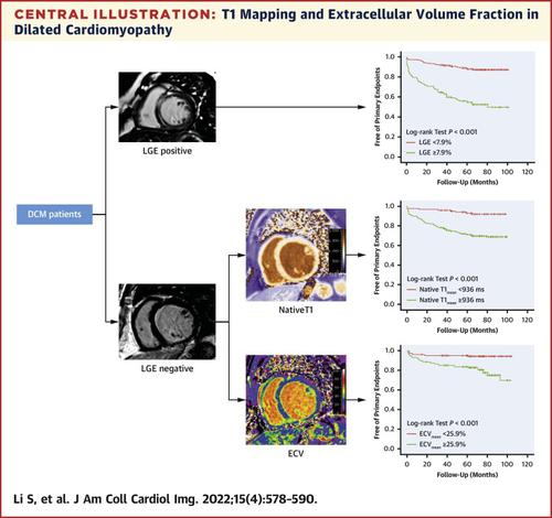
A total of 659 consecutive patients with DCM (498 men; 45 ± 15 years) who underwent cardiac magnetic resonance with T1 mapping and late gadolinium enhancement (LGE) imaging with a 1.5-T magnetic resonance scanner were enrolled in this study. Primary endpoints were cardiac-related death and heart transplantation. Secondary endpoints were hospitalization for heart failure, ventricular arrhythmias, and implantable cardioverter-defibrillator or cardiac resynchronization therapy implantation. Survival estimates were calculated by Kaplan-Meier curves with the log-rank test.
Results
During a mean follow-up of 66.3 ± 20.9 months, 122 and 205 patients with DCM reached the primary and secondary endpoints, respectively. The presence of LGE had an association with both of the primary and secondary endpoints observed in the patients with DCM (both P < 0.001). The maximum native T1 (HR: 1.04; 95% CI: 1.02-1.09) and maximum ECV fraction (HR: 1.14; 95% CI: 1.08-1.21) had associations with the primary endpoints in the patients with positive LGE (both P < 0.001), whereas the mean native T1 (HR: 1.13; 95% CI: 1.10-1.36) and mean ECV fraction (HR: 1.32; 95% CI: 1.12-1.53) had the best associations in the patients with negative LGE (all P < 0.001).
Conclusions
T1 mapping and the ECV fraction had prognostic value in patients with DCM and were particularly important in patients with DCM without LGE. Using a combination of T1 mapping, ECV fraction, and LGE provided optimal risk stratification for patients with DCM.
中文翻译:
扩张型心肌病的 T1 映射和细胞外体积分数
目标
本研究的目的是检查 T1 映射和细胞外体积 (ECV) 分数对扩张型心肌病 (DCM) 患者的预后价值。
背景
具有功能性左心室重构的 DCM 患者预后较差。使用 T1 标测和 ECV 分数对心肌纤维化进行无创评估可能会改善 DCM 患者的风险分层;但是,尚未对此进行系统评估。
方法
共有 659 名 DCM 患者(498 名男性;45 ± 15 岁)接受了 T1 标测的心脏磁共振和使用 1.5-T 磁共振扫描仪进行的晚期钆增强 (LGE) 成像。主要终点是心脏相关死亡和心脏移植。次要终点是因心力衰竭、室性心律失常和植入式心脏复律除颤器或心脏再同步治疗植入而住院。使用对数秩检验通过 Kaplan-Meier 曲线计算生存估计值。
结果
在 66.3 ± 20.9 个月的平均随访期间,122 名和 205 名 DCM 患者分别达到了主要和次要终点。LGE 的存在与在 DCM 患者中观察到的主要和次要终点都有关联(均P < 0.001)。最大天然 T1(HR:1.04;95% CI:1.02-1.09)和最大 ECV 分数(HR:1.14;95% CI:1.08-1.21)与 LGE 阳性患者的主要终点相关(均P < 0.001),而平均天然 T1(HR:1.13;95% CI:1.10-1.36)和平均 ECV 分数(HR:1.32;95% CI:1.12-1.53)在 LGE 阴性患者中具有最佳关联(所有P < 0.001)。
结论
T1 标测和 ECV 分数在 DCM 患者中具有预后价值,对于没有 LGE 的 DCM 患者尤为重要。使用 T1 映射、ECV 分数和 LGE 的组合为 DCM 患者提供了最佳风险分层。


 京公网安备 11010802027423号