Dental Materials ( IF 4.6 ) Pub Date : 2021-09-20 , DOI: 10.1016/j.dental.2021.09.002 Nathanael Leung 1 , Robert A Harper 2 , Bin Zhu 1 , Richard M Shelton 2 , Gabriel Landini 2 , Tan Sui 1
|
Objective
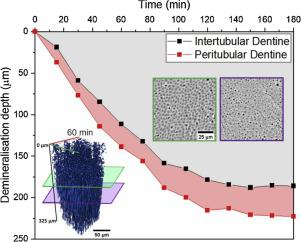
Dental erosion is a common oral condition caused by chronic exposure to acids from intrinsic/extrinsic sources. Repeated acid exposure can lead to the irreversible loss of dental hard tissues (enamel, dentine, cementum). Dentine can become exposed to acid following severe enamel erosion, crown fracture, or gingival recession. Causing hypersensitivity, poor aesthetics, and potential pulp involvement. Improving treatments that can restore the structural integrity and aesthetics are therefore highly desirable. Such developments require a good understanding of how acid demineralisation progresses where relatively little is known in terms of intertubular dentine (ITD) and peritubular dentine (PTD) microstructure. To obtain further insight, this study proposes a new in vitro method for performing demineralisation studies of dentine.
Methods
Advanced high-speed synchrotron X-ray microtomography (SXM), with high spatial (0.325 μm) and temporal (15 min) resolution, was used to conduct the first in vitro, time-resolved 3D (4D) study of the microstructural changes in the ITD and PTD phases of human dentine samples (∼0.8 × 0.8 × 5 mm) during 6 h of continuous acid exposure.
Results
Different demineralisation rates of ITD (1.79 μm/min) and PTD (1.94 μm/min) and their progressive width-depth profiles were quantified, which provide insight for understanding the mechanisms of dentine demineralisation.
Significance
Insights obtained from morphological characterisations and the demineralisation process of ITD and PTD during acid demineralisation would help understand the demineralisation process and potentially aid in developing new therapeutic dentine treatments. This method enables continuous examination of relatively large volumes of dentine during demineralisation and also demonstrates the potential for studying the remineralisation process of proposed therapeutic dentine treatments.
中文翻译:
酸脱矿过程中牙本质小管的 4D 显微结构变化
客观的
牙齿侵蚀是一种常见的口腔疾病,由长期暴露于内在/外在来源的酸引起。反复接触酸会导致牙齿硬组织(牙釉质、牙本质、牙骨质)的不可逆损失。在严重的牙釉质侵蚀、牙冠断裂或牙龈萎缩后,牙本质会暴露在酸中。导致过敏、不美观和潜在的牙髓受累。因此,非常需要改进可以恢复结构完整性和美观的处理方法。这种发展需要很好地了解酸脱矿是如何进行的,而在管间牙本质 (ITD) 和管周牙本质 (PTD) 微观结构方面知之甚少。为了获得更深入的了解,本研究提出了一种新的体外方法来进行牙本质脱矿研究。
方法
先进的高速同步加速器 X 射线显微断层扫描 (SXM),具有高空间 (0.325 μm) 和时间 (15 分钟) 分辨率,用于进行首次体外时间分辨 3D (4D) 研究在连续酸暴露 6 小时期间人类牙本质样品的 ITD 和 PTD 相(~0.8 × 0.8 × 5 mm)。
结果
量化了 ITD (1.79 μm/min) 和 PTD (1.94 μm/min) 的不同脱矿速率及其渐进的宽度-深度曲线,这为理解牙本质脱矿机制提供了见解。
意义
从形态特征以及酸脱矿过程中 ITD 和 PTD 的脱矿过程中获得的见解将有助于了解脱矿过程,并可能有助于开发新的牙本质治疗方法。这种方法能够在脱矿质过程中连续检查相对大量的牙本质,也证明了研究拟议治疗性牙本质治疗的再矿化过程的潜力。


 京公网安备 11010802027423号