当前位置:
X-MOL 学术
›
J. Neurochem.
›
论文详情
Our official English website, www.x-mol.net, welcomes your
feedback! (Note: you will need to create a separate account there.)
The role of neuroimaging in Parkinson’s disease
Journal of Neurochemistry ( IF 4.2 ) Pub Date : 2021-09-17 , DOI: 10.1111/jnc.15516 Natasha S R Bidesi 1 , Ida Vang Andersen 1 , Albert D Windhorst 2 , Vladimir Shalgunov 1 , Matthias M Herth 1, 3
Journal of Neurochemistry ( IF 4.2 ) Pub Date : 2021-09-17 , DOI: 10.1111/jnc.15516 Natasha S R Bidesi 1 , Ida Vang Andersen 1 , Albert D Windhorst 2 , Vladimir Shalgunov 1 , Matthias M Herth 1, 3
Affiliation
|
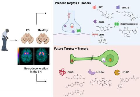
Parkinson's disease (PD) is a neurodegenerative disorder that affects millions of people worldwide. Two hallmarks of PD are the accumulation of alpha-synuclein and the loss of dopaminergic neurons in the brain. There is no cure for PD, and all existing treatments focus on alleviating the symptoms. PD diagnosis is also based on the symptoms, such as abnormalities of movement, mood, and cognition observed in the patients. Molecular imaging methods such as magnetic resonance imaging (MRI), single-photon emission computed tomography (SPECT), and positron emission tomography (PET) can detect objective alterations in the neurochemical machinery of the brain and help diagnose and study neurodegenerative diseases. This review addresses the application of functional MRI, PET, and SPECT in PD patients. We provide an overview of the imaging targets, discuss the rationale behind target selection, the agents (tracers) with which the imaging can be performed, and the main findings regarding each target's state in PD. Molecular imaging has proven itself effective in supporting clinical diagnosis of PD and has helped reveal that PD is a heterogeneous disorder, which has important implications for the development of future therapies. However, the application of molecular imaging for early diagnosis of PD or for differentiation between PD and atypical parkinsonisms has remained challenging. The final section of the review is dedicated to new imaging targets with which one can detect the PD-related pathological changes upstream from dopaminergic degeneration. The foremost of those targets is alpha-synuclein. We discuss the progress of tracer development achieved so far and challenges on the path toward alpha-synuclein imaging in humans.
中文翻译:

神经影像学在帕金森病中的作用
帕金森病 (PD) 是一种影响全球数百万人的神经退行性疾病。PD 的两个标志是 α-突触核蛋白的积累和大脑中多巴胺能神经元的损失。PD 无法治愈,所有现有的治疗都集中在缓解症状上。PD的诊断还基于症状,例如在患者中观察到的运动、情绪和认知异常。分子成像方法,如磁共振成像 (MRI)、单光子发射计算机断层扫描 (SPECT) 和正电子发射断层扫描 (PET),可以检测大脑神经化学机制的客观变化,并帮助诊断和研究神经退行性疾病。本综述探讨了功能性 MRI、PET 和 SPECT 在 PD 患者中的应用。我们提供成像目标的概述,讨论目标选择背后的基本原理、可以进行成像的试剂(示踪剂)以及关于每个目标在 PD 中的状态的主要发现。分子成像已证明其在支持 PD 临床诊断方面是有效的,并有助于揭示 PD 是一种异质性疾病,这对未来疗法的发展具有重要意义。然而,分子影像学在早期诊断 PD 或区分 PD 和非典型帕金森病方面的应用仍然具有挑战性。审查的最后一部分致力于新的成像目标,人们可以利用这些目标检测多巴胺能变性上游的 PD 相关病理变化。这些目标中最重要的是α-突触核蛋白。
更新日期:2021-11-12
中文翻译:
神经影像学在帕金森病中的作用
帕金森病 (PD) 是一种影响全球数百万人的神经退行性疾病。PD 的两个标志是 α-突触核蛋白的积累和大脑中多巴胺能神经元的损失。PD 无法治愈,所有现有的治疗都集中在缓解症状上。PD的诊断还基于症状,例如在患者中观察到的运动、情绪和认知异常。分子成像方法,如磁共振成像 (MRI)、单光子发射计算机断层扫描 (SPECT) 和正电子发射断层扫描 (PET),可以检测大脑神经化学机制的客观变化,并帮助诊断和研究神经退行性疾病。本综述探讨了功能性 MRI、PET 和 SPECT 在 PD 患者中的应用。我们提供成像目标的概述,讨论目标选择背后的基本原理、可以进行成像的试剂(示踪剂)以及关于每个目标在 PD 中的状态的主要发现。分子成像已证明其在支持 PD 临床诊断方面是有效的,并有助于揭示 PD 是一种异质性疾病,这对未来疗法的发展具有重要意义。然而,分子影像学在早期诊断 PD 或区分 PD 和非典型帕金森病方面的应用仍然具有挑战性。审查的最后一部分致力于新的成像目标,人们可以利用这些目标检测多巴胺能变性上游的 PD 相关病理变化。这些目标中最重要的是α-突触核蛋白。


 京公网安备 11010802027423号