当前位置:
X-MOL 学术
›
ACS Appl. Mater. Interfaces
›
论文详情
Our official English website, www.x-mol.net, welcomes your
feedback! (Note: you will need to create a separate account there.)
Ratiometric Fluorescence Imaging of Intracellular MicroRNA with NIR-Assisted Signal Amplification by a Ru-SiO2@Polydopamine Nanoplatform
ACS Applied Materials & Interfaces ( IF 8.3 ) Pub Date : 2021-09-15 , DOI: 10.1021/acsami.1c11324 Xunxun Deng 1, 2 , Xiaobo Liu 1 , Shuo Wu 1 , Shiyu Zang 1 , Xiaotong Lin 1 , Yanqiu Zhao 1 , Chunying Duan 2
ACS Applied Materials & Interfaces ( IF 8.3 ) Pub Date : 2021-09-15 , DOI: 10.1021/acsami.1c11324 Xunxun Deng 1, 2 , Xiaobo Liu 1 , Shuo Wu 1 , Shiyu Zang 1 , Xiaotong Lin 1 , Yanqiu Zhao 1 , Chunying Duan 2
Affiliation
|
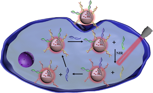
Accurate and sensitive fluorescence imaging of intracellular miRNA is essential for understanding the mechanism underlying some physiological and pathological events, as well as the prevention and diagnosis of diseases. Herein, a highly sensitive ratiometric fluorescent nanoprobe for intracellular miRNA imaging was fabricated by integrating a Ru-SiO2@polydopamine (Ru-SiO2@PDA) nanoplatform with a near-infrared light (NIR)-assisted DNA strand displacement signal amplification strategy. The Ru-SiO2@PDA spheres have excellent biosafety, high photothermal effect, and unique photophysical properties that can both emit a stable red fluorescence and well quench the fluorophores getting closer to them. So, when the fuel DNA and carboxyfluorescein (FAM)-labeled signal DNA are co-assembled on their outer surfaces, the FAM’s green fluorescence is quenched, and a low ratiometric signal is obtained. However, in the presence of miRNA, the target displaces the signal DNA from the capture DNA, releasing the signal DNA far away from the Ru-SiO2@PDA. Then, the green fluorescence recovers and leads to an enhanced Igreen/Ired value. Under NIR light irradiation, the Ru-SiO2@PDA increases the local temperature around the probe and triggers the release of fuel DNA, which thus recycles the target miRNA and effectively amplifies the ratiometric signal. Using A549 cells as a model, the nanoprobe realizes the highly sensitive ratiometric fluorescence imaging of miRNA let-7a, as well as its in vivo up- and down-regulation expressions. It provides a facile tool for highly sensitive and accurate intracellular miRNA detection through one-step incubation and may pave a new avenue for single-cell analysis.
中文翻译:

Ru-SiO2@Polydopamine 纳米平台通过 NIR 辅助信号放大对细胞内 MicroRNA 进行比率荧光成像
细胞内 miRNA 的准确和灵敏的荧光成像对于了解一些生理和病理事件的机制以及疾病的预防和诊断至关重要。在此,通过将 Ru-SiO 2 @polydopamine (Ru-SiO 2 @PDA) 纳米平台与近红外光 (NIR) 辅助 DNA 链置换信号放大策略相结合,制备了一种用于细胞内 miRNA 成像的高灵敏度比率荧光纳米探针。Ru-SiO 2@PDA 球体具有优异的生物安全性、高光热效应和独特的光物理特性,既能发出稳定的红色荧光,又能很好地淬灭靠近它们的荧光团。因此,当燃料 DNA 和羧基荧光素 (FAM) 标记的信号 DNA 在它们的外表面上共同组装时,FAM 的绿色荧光被淬灭,并获得低比率信号。然而,在 miRNA 存在的情况下,靶标从捕获 DNA 中置换出信号 DNA,将信号 DNA 释放到远离 Ru-SiO 2 @PDA 的位置。然后,绿色荧光恢复并导致增强的I green / I red值。在近红外光照射下,Ru-SiO 2@PDA 提高探针周围的局部温度并触发燃料 DNA 的释放,从而回收目标 miRNA 并有效放大比率信号。该纳米探针以A549细胞为模型,实现了miRNA let-7a的高灵敏度比率荧光成像,以及其在体内的上调和下调表达。它通过一步孵育为高灵敏度和准确的细胞内 miRNA 检测提供了一种简便的工具,并可能为单细胞分析开辟一条新途径。
更新日期:2021-09-29
中文翻译:
Ru-SiO2@Polydopamine 纳米平台通过 NIR 辅助信号放大对细胞内 MicroRNA 进行比率荧光成像
细胞内 miRNA 的准确和灵敏的荧光成像对于了解一些生理和病理事件的机制以及疾病的预防和诊断至关重要。在此,通过将 Ru-SiO 2 @polydopamine (Ru-SiO 2 @PDA) 纳米平台与近红外光 (NIR) 辅助 DNA 链置换信号放大策略相结合,制备了一种用于细胞内 miRNA 成像的高灵敏度比率荧光纳米探针。Ru-SiO 2@PDA 球体具有优异的生物安全性、高光热效应和独特的光物理特性,既能发出稳定的红色荧光,又能很好地淬灭靠近它们的荧光团。因此,当燃料 DNA 和羧基荧光素 (FAM) 标记的信号 DNA 在它们的外表面上共同组装时,FAM 的绿色荧光被淬灭,并获得低比率信号。然而,在 miRNA 存在的情况下,靶标从捕获 DNA 中置换出信号 DNA,将信号 DNA 释放到远离 Ru-SiO 2 @PDA 的位置。然后,绿色荧光恢复并导致增强的I green / I red值。在近红外光照射下,Ru-SiO 2@PDA 提高探针周围的局部温度并触发燃料 DNA 的释放,从而回收目标 miRNA 并有效放大比率信号。该纳米探针以A549细胞为模型,实现了miRNA let-7a的高灵敏度比率荧光成像,以及其在体内的上调和下调表达。它通过一步孵育为高灵敏度和准确的细胞内 miRNA 检测提供了一种简便的工具,并可能为单细胞分析开辟一条新途径。


 京公网安备 11010802027423号