Journal of Molecular and Cellular Cardiology ( IF 4.9 ) Pub Date : 2021-09-16 , DOI: 10.1016/j.yjmcc.2021.09.004 Yongyun Li 1 , Jie Yang 1 , Yazhuo Huang 1 , Shengfang Ge 1 , Xin Song 1 , Renbing Jia 1 , Yefei Wang 1
|
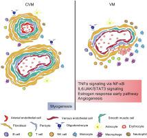
Venous malformation (VM) and cavernous venous malformation (CVM) are two types of vascular malformations. Even if the two diseases are similar in appearance and imaging, the distinct cellular components and signaling pathways between them might help distinguish the two from a molecular perspective. Here, we performed single-cell profiling of 35,245 cells from two VM samples and three CVM samples, with a focus on endothelial cells (ECs), smooth muscle cells (SMCs) and immune microenvironment (IME). Clustering analysis based on differential gene expression unveiled 11 specific cell types, and determined CVM had more SMCs. Re-clustering of ECs and SMCs indicated CVM was dominated by arterial components, while VM is dominated by venous components. Gene set variation analysis suggested the activation of inflammation-related pathways in VM ECs, and upregulation of myogenesis pathway in CVM SMCs. In IME analysis, immune cells were identified to accounted for nearly 30% of the total cell number, including macrophages, monocytes, NK cells, T cells and B cells. Notably, more macrophages and monocytes were discovered in VM, indicating innate immune responses might be more closely related to VM pathogenesis. In addition, angiogenesis pathway was highlighted among the significant pathways of macrophages & monocytes between CVM and VM. In VM, VEGFA was highly expressed in macrophages & monocytes, while its receptors were all abundantly present in ECs. The close interaction of VEGFA on macrophages with its receptors on ECs was also predicted by CellPhoneDB analysis. Our results document cellular composition, significant pathways, and critical IME in CVM and VM development.
中文翻译:
单细胞转录组揭示静脉畸形和海绵状静脉畸形中的细胞异质性和免疫微环境
静脉畸形(VM)和海绵状静脉畸形(CVM)是两种类型的血管畸形。即使这两种疾病在外观和影像学上相似,但它们之间不同的细胞成分和信号通路可能有助于从分子角度区分两者。在这里,我们对来自两个 VM 样本和三个 CVM 样本的 35,245 个细胞进行了单细胞分析,重点是内皮细胞 (EC)、平滑肌细胞 (SMC) 和免疫微环境 (IME)。基于差异基因表达的聚类分析揭示了 11 种特定细胞类型,并确定 CVM 具有更多的 SMC。ECs 和 SMCs 的重新聚类表明 CVM 以动脉成分为主,而 VM 以静脉成分为主。基因组变异分析表明 VM EC 中炎症相关通路的激活,和 CVM SMC 中肌生成途径的上调。在IME分析中,免疫细胞被确定占细胞总数的近30%,包括巨噬细胞、单核细胞、NK细胞、T细胞和B细胞。值得注意的是,在 VM 中发现了更多的巨噬细胞和单核细胞,表明先天免疫反应可能与 VM 发病机制更密切相关。此外,在CVM和VM之间的巨噬细胞和单核细胞的重要途径中,血管生成途径被突出显示。在VM中,VEGFA在巨噬细胞和单核细胞中高表达,而其受体在ECs中都大量存在。CellPhoneDB分析还预测了巨噬细胞上的VEGFA与其EC上的受体的密切相互作用。我们的结果记录了 CVM 和 VM 开发中的细胞组成、重要途径和关键 IME。


 京公网安备 11010802027423号