Bioactive Materials ( IF 18.0 ) Pub Date : 2021-09-16 , DOI: 10.1016/j.bioactmat.2021.09.018 Yunfan Xue 1 , Honglin Qian 1 , Xu Li 1 , Jing Wang 1 , Kefeng Ren 1 , Jian Ji 1
|
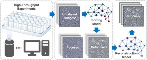
The increasing throughput of experiments in biomaterials research makes automatic techniques more and more necessary. Among all the characterization methods, microscopy makes fundamental contributions to biomaterials science where precisely focused images are the basis of related research. Although automatic focusing has been widely applied in all kinds of microscopes, defocused images can still be acquired now and then due to factors including background noises of materials and mechanical errors. Herein, we present a deep-learning-based method for the automatic sorting and reconstruction of defocused cell images. First, the defocusing problem is illustrated on a high-throughput cell microarray. Then, a comprehensive dataset of phase-contrast images captured from varied conditions containing multiple cell types, magnifications, and substrate materials is prepared to establish and test our method. We obtain high accuracy of over 0.993 on the dataset using a simple network architecture that requires less than half of the training time compared with the classical ResNetV2 architecture. Moreover, the subcellular-level reconstruction of heavily defocused cell images is achieved with another architecture. The applicability of the established workflow in practice is finally demonstrated on the high-throughput cell microarray. The intelligent workflow does not require a priori knowledge of focusing algorithms, possessing widespread application value in cell experiments concerning high-throughput or time-lapse imaging.
中文翻译:
一种基于深度学习的工作流程,用于处理高通量实验中的散焦问题
生物材料研究中实验量的增加使得自动化技术越来越必要。在所有表征方法中,显微镜对生物材料科学做出了根本性的贡献,其中精确聚焦的图像是相关研究的基础。尽管自动对焦已广泛应用于各种显微镜,但由于材料的背景噪声和机械误差等因素,有时仍会出现散焦图像。在此,我们提出了一种基于深度学习的方法,用于散焦细胞图像的自动排序和重建。首先,在高通量细胞微阵列上说明散焦问题。然后,从包含多种细胞类型、放大倍数、准备好基板材料以建立和测试我们的方法。我们使用简单的网络架构在数据集上获得了超过 0.993 的高精度,与经典的 ResNetV2 架构相比,该架构所需的训练时间不到一半。此外,严重散焦细胞图像的亚细胞级重建是通过另一种架构实现的。已建立的工作流程在实践中的适用性最终在高通量细胞微阵列上得到证明。智能工作流程不需要聚焦算法的先验知识,在高通量或延时成像的细胞实验中具有广泛的应用价值。993 在数据集上使用简单的网络架构,与经典的 ResNetV2 架构相比,所需的训练时间不到一半。此外,严重散焦细胞图像的亚细胞级重建是通过另一种架构实现的。已建立的工作流程在实践中的适用性最终在高通量细胞微阵列上得到证明。智能工作流程不需要聚焦算法的先验知识,在高通量或延时成像的细胞实验中具有广泛的应用价值。993 在数据集上使用简单的网络架构,与经典的 ResNetV2 架构相比,所需的训练时间不到一半。此外,严重散焦细胞图像的亚细胞级重建是通过另一种架构实现的。已建立的工作流程在实践中的适用性最终在高通量细胞微阵列上得到证明。智能工作流程不需要聚焦算法的先验知识,在高通量或延时成像的细胞实验中具有广泛的应用价值。已建立的工作流程在实践中的适用性最终在高通量细胞微阵列上得到证明。智能工作流程不需要聚焦算法的先验知识,在高通量或延时成像的细胞实验中具有广泛的应用价值。已建立的工作流程在实践中的适用性最终在高通量细胞微阵列上得到证明。智能工作流程不需要聚焦算法的先验知识,在高通量或延时成像的细胞实验中具有广泛的应用价值。


 京公网安备 11010802027423号