Cell Reports ( IF 7.5 ) Pub Date : 2021-09-14 , DOI: 10.1016/j.celrep.2021.109707 Ke Yang 1 , Min Liu 1 , Zhi Feng 1 , Marta Rojas 2 , Lingjian Zhou 1 , Hongmei Ke 1 , José Carlos Pastor-Pareja 3
|
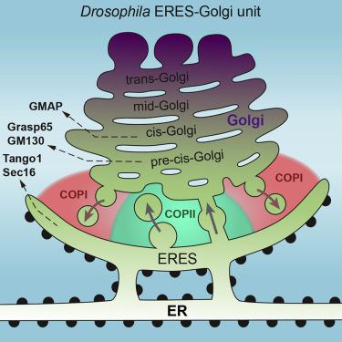
Secretory cargos are collected at endoplasmic reticulum (ER) exit sites (ERES) before transport to the Golgi apparatus. Decades of research have provided many details of the molecular events underlying ER-Golgi exchanges. Essential questions, however, remain about the organization of the ER-Golgi interface in cells and the type of membrane structures mediating traffic from ERES. To investigate these, we use transgenic tagging in Drosophila flies, 3D-structured illumination microscopy (SIM), and focused ion beam scanning electron microscopy (FIB-SEM) to characterize ERES-Golgi units in collagen-producing fat body, imaginal discs, and imaginal discs overexpressing ERES determinant Tango1. Facing ERES, we find a pre-cis-Golgi region, equivalent to the vertebrate ER-Golgi intermediate compartment (ERGIC), involved in both anterograde and retrograde transport. This pre-cis-Golgi is continuous with the rest of the Golgi, not a separate compartment or collection of large carriers, for which we find no evidence. We observe, however, many vesicles, as well as pearled tubules connecting ERES and Golgi.
中文翻译:
果蝇 ER 出口位点显示丰富的 ER-高尔基体囊泡和珠状管,但没有巨型载体
在运输到高尔基体之前,在内质网 (ER) 出口部位 (ERES) 收集分泌物。数十年的研究提供了许多关于 ER-高尔基体交换的分子事件的细节。然而,基本问题仍然是关于细胞中 ER-高尔基界面的组织以及介导 ERES 流量的膜结构类型。为了研究这些,我们在果蝇中使用转基因标记、3D 结构照明显微镜 (SIM) 和聚焦离子束扫描电子显微镜 (FIB-SEM) 来表征产生胶原蛋白的脂肪体、成像盘和过度表达 ERES 行列式 Tango1 的成像盘。面对 ERES,我们找到了一个精确的-Golgi 区域,相当于脊椎动物 ER-Golgi 中间隔室 (ERGIC),参与顺行和逆行运输。这个精确的高尔基体与高尔基体的其余部分是连续的,而不是一个单独的隔间或大型载体的集合,我们找不到任何证据。然而,我们观察到许多囊泡,以及连接 ERES 和高尔基体的珠状小管。


 京公网安备 11010802027423号