当前位置:
X-MOL 学术
›
J. Neurosci. Res.
›
论文详情
Our official English website, www.x-mol.net, welcomes your
feedback! (Note: you will need to create a separate account there.)
Diffusion tensor imaging-based pontine damage as a degeneration marker in synucleinopathy
Journal of Neuroscience Research ( IF 2.9 ) Pub Date : 2021-09-14 , DOI: 10.1002/jnr.24926 Seok Jong Chung 1, 2 , Kyoo Ho Cho 1, 3 , Yang Hyun Lee 1 , Han Soo Yoo 1 , KyoungWon Baik 1 , Jin Ho Jung 1, 4 , Byoung Seok Ye 1 , Young H Sohn 1 , Jungho Cha 5 , Phil Hyu Lee 1, 6
Journal of Neuroscience Research ( IF 2.9 ) Pub Date : 2021-09-14 , DOI: 10.1002/jnr.24926 Seok Jong Chung 1, 2 , Kyoo Ho Cho 1, 3 , Yang Hyun Lee 1 , Han Soo Yoo 1 , KyoungWon Baik 1 , Jin Ho Jung 1, 4 , Byoung Seok Ye 1 , Young H Sohn 1 , Jungho Cha 5 , Phil Hyu Lee 1, 6
Affiliation
|
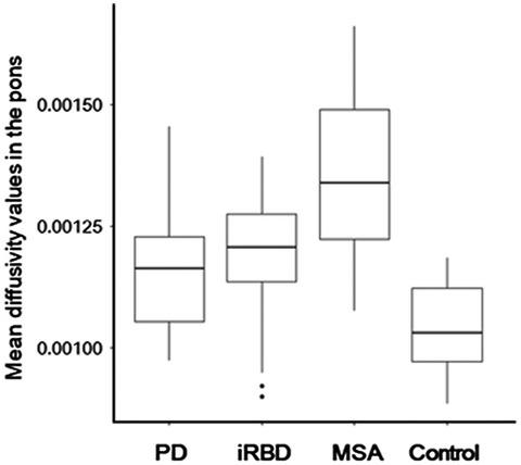
The pons is one of the earliest affected regions in patients with synucleinopathies. We aimed to investigate the diagnostic value of measuring pontine damage using diffusion tensor imaging (DTI) in these patients. We enrolled 49 patients with Parkinson's disease (PD), 16 patients with idiopathic rapid eye movement sleep behavior disorder (iRBD), 23 patients with multiple system atrophy (MSA), and 39 healthy controls in this study. All the participants underwent high-resolution T1-weighted imaging and DTI. Mean diffusivity (MD) and fraction anisotropy (FA) values in the pons were calculated to characterize structural damage. The discriminatory power of pontine MD and FA values to differentiate patients with synucleinopathies from healthy controls was examined using receiver operating characteristics (ROC) analyses. Compared to healthy controls, patients with PD, iRBD, and MSA had increased MD values and decreased FA values in the pons, although no correlation was observed between these DTI measures and disease severity. The ROC analyses showed that MD values in the pons had a fair discriminatory power to differentiate healthy controls from patients with PD (area under the curve [AUC], 0.813), iRBD (AUC, 0.779), and MSA (AUC, 0.951). The AUC for pontine FA values was smaller than that for pontine MD values when differentiating healthy controls from patients with PD (AUC, 0.713; p = 0.054) and iRBD (AUC, 0.686; p = 0.045). Our results suggest that MD values in the pons may be a useful marker of brain stem neurodegeneration in patients with synucleinopathies.
中文翻译:

基于扩散张量成像的脑桥损伤作为突触核蛋白病的退化标志物
脑桥是突触核蛋白病患者最早受影响的区域之一。我们旨在研究使用弥散张量成像 (DTI) 测量这些患者的脑桥损伤的诊断价值。我们在本研究中招募了 49 名帕金森病 (PD) 患者、16 名特发性快速眼动睡眠行为障碍 (iRBD) 患者、23 名多系统萎缩 (MSA) 患者和 39 名健康对照。所有参与者都接受了高分辨率 T1 加权成像和 DTI。计算脑桥中的平均扩散率 (MD) 和分数各向异性 (FA) 值来表征结构损伤。使用接受者操作特征 (ROC) 分析检查 pontine MD 和 FA 值区分突触核蛋白病患者与健康对照的区分能力。与健康对照组相比,PD、iRBD 和 MSA 患者的脑桥 MD 值增加而 FA 值降低,尽管在这些 DTI 测量值与疾病严重程度之间没有观察到相关性。ROC 分析表明,脑桥中的 MD 值具有公平的区分能力,可以区分健康对照与 PD(曲线下面积 [AUC],0.813)、iRBD(AUC,0.779)和 MSA(AUC,0.951)患者。在区分健康对照和 PD 患者时,脑桥 FA 值的 AUC 小于脑桥 MD 值(AUC,0.713;ROC 分析表明,脑桥中的 MD 值具有公平的区分能力,可以区分健康对照与 PD(曲线下面积 [AUC],0.813)、iRBD(AUC,0.779)和 MSA(AUC,0.951)患者。在区分健康对照和 PD 患者时,脑桥 FA 值的 AUC 小于脑桥 MD 值(AUC,0.713;ROC 分析表明,脑桥中的 MD 值具有公平的区分能力,可以区分健康对照与 PD(曲线下面积 [AUC],0.813)、iRBD(AUC,0.779)和 MSA(AUC,0.951)患者。在区分健康对照和 PD 患者时,脑桥 FA 值的 AUC 小于脑桥 MD 值(AUC,0.713;p = 0.054)和 iRBD(AUC,0.686;p = 0.045)。我们的研究结果表明,脑桥中的 MD 值可能是突触核蛋白病患者脑干神经变性的有用标志物。
更新日期:2021-09-14
中文翻译:
基于扩散张量成像的脑桥损伤作为突触核蛋白病的退化标志物
脑桥是突触核蛋白病患者最早受影响的区域之一。我们旨在研究使用弥散张量成像 (DTI) 测量这些患者的脑桥损伤的诊断价值。我们在本研究中招募了 49 名帕金森病 (PD) 患者、16 名特发性快速眼动睡眠行为障碍 (iRBD) 患者、23 名多系统萎缩 (MSA) 患者和 39 名健康对照。所有参与者都接受了高分辨率 T1 加权成像和 DTI。计算脑桥中的平均扩散率 (MD) 和分数各向异性 (FA) 值来表征结构损伤。使用接受者操作特征 (ROC) 分析检查 pontine MD 和 FA 值区分突触核蛋白病患者与健康对照的区分能力。与健康对照组相比,PD、iRBD 和 MSA 患者的脑桥 MD 值增加而 FA 值降低,尽管在这些 DTI 测量值与疾病严重程度之间没有观察到相关性。ROC 分析表明,脑桥中的 MD 值具有公平的区分能力,可以区分健康对照与 PD(曲线下面积 [AUC],0.813)、iRBD(AUC,0.779)和 MSA(AUC,0.951)患者。在区分健康对照和 PD 患者时,脑桥 FA 值的 AUC 小于脑桥 MD 值(AUC,0.713;ROC 分析表明,脑桥中的 MD 值具有公平的区分能力,可以区分健康对照与 PD(曲线下面积 [AUC],0.813)、iRBD(AUC,0.779)和 MSA(AUC,0.951)患者。在区分健康对照和 PD 患者时,脑桥 FA 值的 AUC 小于脑桥 MD 值(AUC,0.713;ROC 分析表明,脑桥中的 MD 值具有公平的区分能力,可以区分健康对照与 PD(曲线下面积 [AUC],0.813)、iRBD(AUC,0.779)和 MSA(AUC,0.951)患者。在区分健康对照和 PD 患者时,脑桥 FA 值的 AUC 小于脑桥 MD 值(AUC,0.713;p = 0.054)和 iRBD(AUC,0.686;p = 0.045)。我们的研究结果表明,脑桥中的 MD 值可能是突触核蛋白病患者脑干神经变性的有用标志物。


 京公网安备 11010802027423号