Journal of Materials Science: Materials in Medicine ( IF 3.7 ) Pub Date : 2021-09-15 , DOI: 10.1007/s10856-021-06600-z Zhengye Zhang 1 , Yang Zheng 1 , Jianing Zu 1 , Jinpeng Zhuang 1 , Gongping Xu 1 , Jinglong Yan 1 , Xiaoqi Liu 1
|
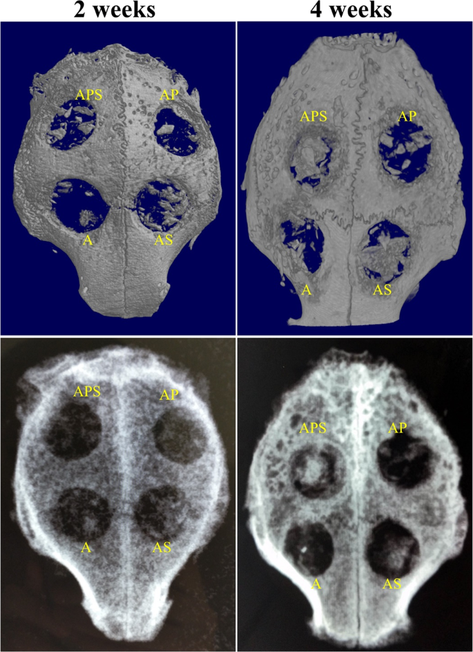
The current study aimed to evaluate the effects of chemokine stromal cell-derived factor (SDF)-1α and platelet-rich plasma (PRP) on bone formation and angiogenesis, and to assess whether SDF-1α and PRP could function synergistically. Four evenly distributed defects (8 mm in diameter) were generated in the calvarial bones of New Zealand white rabbits. All rabbits received four treatment regimens containing autogenous bone particles (AB), SDF-1α, or PRP. AB group presented significantly less bone formation compared with the other three groups 2 and 4 weeks after surgery. The amount of newly formed bone in the AB+PRP+SDF-1α group was similar to that in the AB + SDF-1α group at the 4-week time-point (p = 0.65), and was much greater than that in the AB and AB+PRP group (p < 0.001). Meanwhile, more new blood vessels were formed in the AB+PRP, AB+SDF-1α, and AB+PRP+SDF-1α group versus the AB group. AB+PRP+SDF-1α group showed statistically increased angiogenesis compared with the AB+PRP and AB+SDF-1α groups (both p < 0.05) after treatment for 2 and 4 weeks. These findings indicated that SDF-1α and PRP might exhibit synergistic effects to promote angiogenesis in early bone regeneration.
中文翻译:
基质细胞衍生因子 (SDF)-1α 和富含血小板的血浆在兔颅盖骨原位同时增强骨再生和血管生成
本研究旨在评估趋化因子基质细胞衍生因子 (SDF)-1α 和富血小板血浆 (PRP) 对骨形成和血管生成的影响,并评估 SDF-1α 和 PRP 是否可以协同发挥作用。在新西兰白兔的颅骨中产生了四个均匀分布的缺陷(直径 8 毫米)。所有兔子都接受了四种含有自体骨颗粒 (AB)、SDF-1α 或 PRP 的治疗方案。术后 2 周和 4 周,AB 组的骨形成明显少于其他三组。AB+PRP+SDF-1α组在4周时间点的新生骨量与AB+SDF-1α组相似(p =0.65),远大于对照组。 AB和AB+PRP组(p < 0.001)。同时,与AB组相比,AB+PRP、AB+SDF-1α和AB+PRP+SDF-1α组有更多新生血管形成。与 AB+PRP 和 AB+SDF-1α 组相比,AB+PRP+SDF-1α 组 在治疗 2 周和 4 周后显示出统计学上增加的血管生成(均p < 0.05)。这些发现表明,SDF-1α 和 PRP 可能表现出协同作用,以促进早期骨再生中的血管生成。


 京公网安备 11010802027423号