当前位置:
X-MOL 学术
›
Hum. Brain Mapp.
›
论文详情
Our official English website, www.x-mol.net, welcomes your
feedback! (Note: you will need to create a separate account there.)
Automated claustrum segmentation in human brain MRI using deep learning
Human Brain Mapping ( IF 3.5 ) Pub Date : 2021-09-14 , DOI: 10.1002/hbm.25655 Hongwei Li 1, 2 , Aurore Menegaux 3, 4 , Benita Schmitz-Koep 3, 4 , Antonia Neubauer 3, 4 , Felix J B Bäuerlein 5 , Suprosanna Shit 1 , Christian Sorg 3, 4, 6 , Bjoern Menze 1, 2 , Dennis Hedderich 3, 4
Human Brain Mapping ( IF 3.5 ) Pub Date : 2021-09-14 , DOI: 10.1002/hbm.25655 Hongwei Li 1, 2 , Aurore Menegaux 3, 4 , Benita Schmitz-Koep 3, 4 , Antonia Neubauer 3, 4 , Felix J B Bäuerlein 5 , Suprosanna Shit 1 , Christian Sorg 3, 4, 6 , Bjoern Menze 1, 2 , Dennis Hedderich 3, 4
Affiliation
|
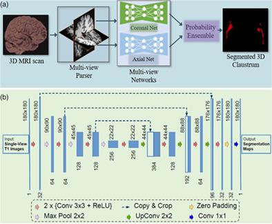
In the last two decades, neuroscience has produced intriguing evidence for a central role of the claustrum in mammalian forebrain structure and function. However, relatively few in vivo studies of the claustrum exist in humans. A reason for this may be the delicate and sheet-like structure of the claustrum lying between the insular cortex and the putamen, which makes it not amenable to conventional segmentation methods. Recently, Deep Learning (DL) based approaches have been successfully introduced for automated segmentation of complex, subcortical brain structures. In the following, we present a multi-view DL-based approach to segment the claustrum in T1-weighted MRI scans. We trained and evaluated the proposed method in 181 individuals, using bilateral manual claustrum annotations by an expert neuroradiologist as reference standard. Cross-validation experiments yielded median volumetric similarity, robust Hausdorff distance, and Dice score of 93.3%, 1.41 mm, and 71.8%, respectively, representing equal or superior segmentation performance compared to human intra-rater reliability. The leave-one-scanner-out evaluation showed good transferability of the algorithm to images from unseen scanners at slightly inferior performance. Furthermore, we found that DL-based claustrum segmentation benefits from multi-view information and requires a sample size of around 75 MRI scans in the training set. We conclude that the developed algorithm allows for robust automated claustrum segmentation and thus yields considerable potential for facilitating MRI-based research of the human claustrum. The software and models of our method are made publicly available.
中文翻译:

使用深度学习在人脑 MRI 中自动进行屏状分割
在过去的二十年里,神经科学已经产生了有趣的证据,证明锁骨在哺乳动物前脑结构和功能中的核心作用。然而,在人类中存在相对较少的柱状体的体内研究。造成这种情况的一个原因可能是位于岛叶皮层和壳核之间的晶状体的精致和片状结构,这使得它不适合传统的分割方法。最近,基于深度学习 (DL) 的方法已成功用于复杂的皮层下大脑结构的自动分割。在下文中,我们提出了一种基于多视图 DL 的方法来分割 T1 加权 MRI 扫描中的锁骨。我们使用专家神经放射学家的双边手动锁骨注释作为参考标准,在 181 个人中对所提出的方法进行了培训和评估。交叉验证实验产生了中值体积相似性、稳健的 Hausdorff 距离和 Dice 得分分别为 93.3%、1.41 mm 和 71.8%,与人类内部评估者可靠性相比,代表了同等或更高的分割性能。留一扫描仪评估表明,该算法对来自看不见的扫描仪的图像具有良好的可迁移性,但性能稍差。此外,我们发现基于 DL 的 claustrum 分割受益于多视图信息,并且需要在训练集中进行大约 75 次 MRI 扫描的样本大小。我们得出的结论是,所开发的算法允许强大的自动锁骨分割,因此为促进基于 MRI 的人类锁骨研究产生了巨大的潜力。我们方法的软件和模型是公开的。
更新日期:2021-11-17
中文翻译:
使用深度学习在人脑 MRI 中自动进行屏状分割
在过去的二十年里,神经科学已经产生了有趣的证据,证明锁骨在哺乳动物前脑结构和功能中的核心作用。然而,在人类中存在相对较少的柱状体的体内研究。造成这种情况的一个原因可能是位于岛叶皮层和壳核之间的晶状体的精致和片状结构,这使得它不适合传统的分割方法。最近,基于深度学习 (DL) 的方法已成功用于复杂的皮层下大脑结构的自动分割。在下文中,我们提出了一种基于多视图 DL 的方法来分割 T1 加权 MRI 扫描中的锁骨。我们使用专家神经放射学家的双边手动锁骨注释作为参考标准,在 181 个人中对所提出的方法进行了培训和评估。交叉验证实验产生了中值体积相似性、稳健的 Hausdorff 距离和 Dice 得分分别为 93.3%、1.41 mm 和 71.8%,与人类内部评估者可靠性相比,代表了同等或更高的分割性能。留一扫描仪评估表明,该算法对来自看不见的扫描仪的图像具有良好的可迁移性,但性能稍差。此外,我们发现基于 DL 的 claustrum 分割受益于多视图信息,并且需要在训练集中进行大约 75 次 MRI 扫描的样本大小。我们得出的结论是,所开发的算法允许强大的自动锁骨分割,因此为促进基于 MRI 的人类锁骨研究产生了巨大的潜力。我们方法的软件和模型是公开的。


 京公网安备 11010802027423号