Journal of Neuroscience Methods ( IF 2.7 ) Pub Date : 2021-09-13 , DOI: 10.1016/j.jneumeth.2021.109354 Griffin Rodgers 1 , Willy Kuo 2 , Georg Schulz 1 , Mario Scheel 3 , Alexandra Migga 1 , Christos Bikis 4 , Christine Tanner 1 , Vartan Kurtcuoglu 2 , Timm Weitkamp 3 , Bert Müller 1
|
Background
Micrometer-resolution neuroimaging with gold-standard conventional histology requires tissue fixation and embedding. The exchange of solvents for the creation of sectionable paraffin blocks modifies tissue density and generates non-uniform brain shrinkage.
New method
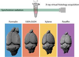
We employed synchrotron radiation-based X-ray microtomography for slicing- and label-free virtual histology of the mouse brain at different stages of the standard preparation protocol from formalin fixation via ascending ethanol solutions and xylene to paraffin embedding. Segmentation of anatomical regions allowed us to quantify non-uniform tissue shrinkage. Global and local changes in X-ray absorption gave insight into contrast enhancement for virtual histology.
Results
The volume of the entire mouse brain was 60%, 56%, and 40% of that in formalin for, respectively, 100% ethanol, xylene, and paraffin. The volume changes of anatomical regions such as the hippocampus, anterior commissure, and ventricles differ from the global volume change. X-ray absorption of the full brain decreased, while local absorption differences increased, resulting in enhanced contrast for virtual histology. These trends were also observed with laboratory microtomography measurements.
Comparison with existing methods
Microtomography provided sub-10 μm spatial resolution with sufficient density resolution to resolve anatomical structures at each step of the embedding protocol. The spatial resolution of conventional computed tomography and magnetic resonance microscopy is an order of magnitude lower and both do not match the contrast of microtomography over the entire embedding protocol. Unlike feature-to-feature or total volume measurements, our approach allows for calculation of volume change based on segmentation.
Conclusion
We present isotropic micrometer-resolution imaging to quantify morphology and composition changes in a mouse brain during the standard histological preparation. The proposed method can be employed to identify the most appropriate embedding medium for anatomical feature visualization, to reveal the basis for the dramatic X-ray contrast enhancement observed in numerous embedded tissues, and to quantify morphological changes during tissue fixation and embedding.
中文翻译:
从福尔马林固定到石蜡包埋的整个小鼠大脑的虚拟组织学。第 1 部分:数据采集、解剖特征分割、跟踪全局体积和密度变化
背景
具有金标准常规组织学的微米分辨率神经成像需要组织固定和嵌入。用于创建可切片石蜡块的溶剂交换会改变组织密度并产生不均匀的大脑收缩。
新方法
我们采用基于同步辐射的 X 射线显微断层扫描,在标准制备方案的不同阶段对小鼠大脑进行切片和无标记虚拟组织学,从福尔马林固定通过升乙醇溶液和二甲苯到石蜡包埋。解剖区域的分割使我们能够量化不均匀的组织收缩。X 射线吸收的全局和局部变化让我们深入了解虚拟组织学的对比度增强。
结果
对于 100% 乙醇、二甲苯和石蜡,整个小鼠大脑的体积分别是福尔马林中体积的 60%、56% 和 40%。海马、前连合和脑室等解剖区域的体积变化与整体体积变化不同。全脑的 X 射线吸收减少,而局部吸收差异增加,导致虚拟组织学的对比度增强。通过实验室显微断层扫描测量也观察到这些趋势。
与现有方法的比较
显微断层扫描提供亚 10 μ m 空间分辨率和足够的密度分辨率,以在嵌入协议的每个步骤解析解剖结构。传统计算机断层扫描和磁共振显微镜的空间分辨率低一个数量级,两者都不匹配整个嵌入协议的显微断层扫描的对比度。与特征到特征或总体积测量不同,我们的方法允许基于分割计算体积变化。
结论
我们提出了各向同性微米分辨率成像,以量化标准组织学制备过程中小鼠大脑的形态和成分变化。所提出的方法可用于确定最适合解剖特征可视化的嵌入介质,揭示在众多嵌入组织中观察到的显着 X 射线对比度增强的基础,并量化组织固定和嵌入过程中的形态变化。


 京公网安备 11010802027423号