iScience ( IF 4.6 ) Pub Date : 2021-09-11 , DOI: 10.1016/j.isci.2021.103127 Jake W Willows 1, 2 , Magdalena Blaszkiewicz 1, 2, 3 , Amy Lamore 4 , Samuel Borer 1 , Amanda L Dubois 4 , Emma Garner 1 , William P Breeding 5 , Karissa B Tilbury 3, 5 , Andre Khalil 3, 5, 6 , Kristy L Townsend 1, 2, 3, 4, 5
|
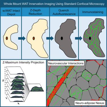
Little is known about the diversity and function of adipose tissue nerves, due in part to the inability to effectively visualize the tissue’s diverse nerve subtypes and the patterns of innervation across an intact depot. The tools to image and quantify adipose tissue innervation are currently limited. Here, we present a method of tissue processing that decreases tissue thickness in the z-axis while leaving cells intact for subsequent immunostaining. This was combined with autofluorescence quenching techniques to permit intact whole tissues to be mounted on slides and imaged by confocal microscopy, with a complementary means to perform whole tissue neurite density quantification after capture of tiled z-stack images. Additionally, we demonstrate how to visualize nerve terminals (the neuro-adipose nexus) in intact blocks of adipose tissue without z-depth reduction. We have included examples of data demonstrating nerve subtypes, neurovascular interactions, label-free imaging of collagen, and nerve bundle digital cross-sections.
中文翻译:
全库脂肪组织神经支配的可视化和分析
人们对脂肪组织神经的多样性和功能知之甚少,部分原因是无法有效地可视化组织的不同神经亚型和完整神经支配的模式。目前对脂肪组织神经支配进行成像和量化的工具有限。在这里,我们提出了一种组织处理方法,可以减少 z 轴上的组织厚度,同时保持细胞完整以进行后续的免疫染色。这与自发荧光猝灭技术相结合,允许将完整的整个组织安装在载玻片上并通过共焦显微镜成像,并在捕获平铺 z 堆栈图像后使用补充方法进行整个组织神经突密度量化。此外,我们演示了如何在不减少 z 深度的情况下可视化完整脂肪组织块中的神经末梢(神经脂肪连接)。我们提供了展示神经亚型、神经血管相互作用、胶原蛋白无标记成像和神经束数字横截面的数据示例。


 京公网安备 11010802027423号