当前位置:
X-MOL 学术
›
Eur. J. Neurosci.
›
论文详情
Our official English website, www.x-mol.net, welcomes your
feedback! (Note: you will need to create a separate account there.)
Dysfunction of oligodendrocyte inwardly rectifying potassium channel in a rat model of amyotrophic lateral sclerosis
European Journal of Neuroscience ( IF 2.7 ) Pub Date : 2021-09-12 , DOI: 10.1111/ejn.15451 Mina Peric 1 , Ljiljana Nikolic 2 , Pavle R Andjus 1 , Danijela Bataveljic 1
European Journal of Neuroscience ( IF 2.7 ) Pub Date : 2021-09-12 , DOI: 10.1111/ejn.15451 Mina Peric 1 , Ljiljana Nikolic 2 , Pavle R Andjus 1 , Danijela Bataveljic 1
Affiliation
|
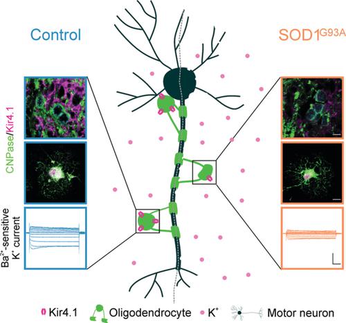
Amyotrophic lateral sclerosis (ALS) is a fatal neurodegenerative disease caused by the death of motor neurons in the spinal cord and the brain. Although this disease is characterized by motoneuron degeneration, non-neuronal cells such as oligodendrocytes play an important role in the disease onset and progression. The aim of our study was to examine functional properties of oligodendrocytes in the SOD1G93A rat model of ALS with a particular focus on the inwardly rectifying potassium channel Kir4.1 that is abundantly expressed in these glial cells and plays a role in the regulation of extracellular K+. First, we demonstrate that the expression of Kir4.1 is diminished in the spinal cord oligodendrocytes of the SOD1G93A rat. Moreover, our data show an elevated number of dysmorphic oligodendrocytes in the ALS spinal cord that is indicative of a degenerative phenotype. In order to assess physiological properties of oligodendrocytes, we prepared cell cultures from the rat spinal cord. Oligodendrocytes isolated from the SOD1G93A spinal cord display similar ramification of the processes as the control but express a lower level of Kir4.1. We further demonstrate an impairment of oligodendrocyte functional properties in ALS. Remarkably, whole-cell patch-clamp recordings revealed compromised membrane biophysical properties and diminished inward currents in the SOD1G93A oligodendrocytes. In addition, the Ba2+-sensitive Kir currents were decreased in ALS oligodendrocytes. Altogether, our findings provide the evidence of impaired Kir4.1 expression and function in oligodendrocytes of the SOD1G93A spinal cord, suggesting oligodendrocyte Kir4.1 channel as a potential contributor to the ALS pathophysiology.
中文翻译:

肌萎缩侧索硬化大鼠模型少突胶质细胞内向整流钾通道功能障碍
肌萎缩侧索硬化症 (ALS) 是一种致命的神经退行性疾病,由脊髓和大脑中的运动神经元死亡引起。尽管这种疾病的特征是运动神经元变性,但非神经元细胞如少突胶质细胞在疾病的发作和进展中起重要作用。我们研究的目的是检查 ALS 的 SOD1 G93A大鼠模型中少突胶质细胞的功能特性,特别关注在这些神经胶质细胞中大量表达并在细胞外调节中发挥作用的内向整流钾通道 Kir4.1 ķ +。首先,我们证明了在 SOD1 G93A的脊髓少突胶质细胞中 Kir4.1 的表达减少鼠。此外,我们的数据显示 ALS 脊髓中畸形少突胶质细胞数量增加,这表明存在退行性表型。为了评估少突胶质细胞的生理特性,我们从大鼠脊髓制备细胞培养物。从 SOD1 G93A脊髓分离的少突胶质细胞显示出与对照相似的过程分支,但表达较低水平的 Kir4.1。我们进一步证明了 ALS 中少突胶质细胞功能特性的损害。值得注意的是,全细胞膜片钳记录显示 SOD1 G93A少突胶质细胞的膜生物物理特性受损,内向电流减少。此外,Ba 2+ALS少突胶质细胞中敏感的Kir电流降低。总之,我们的研究结果提供了 SOD1 G93A脊髓少突胶质细胞中 Kir4.1 表达和功能受损的证据,表明少突胶质细胞 Kir4.1 通道是 ALS 病理生理学的潜在贡献者。
更新日期:2021-10-20
中文翻译:
肌萎缩侧索硬化大鼠模型少突胶质细胞内向整流钾通道功能障碍
肌萎缩侧索硬化症 (ALS) 是一种致命的神经退行性疾病,由脊髓和大脑中的运动神经元死亡引起。尽管这种疾病的特征是运动神经元变性,但非神经元细胞如少突胶质细胞在疾病的发作和进展中起重要作用。我们研究的目的是检查 ALS 的 SOD1 G93A大鼠模型中少突胶质细胞的功能特性,特别关注在这些神经胶质细胞中大量表达并在细胞外调节中发挥作用的内向整流钾通道 Kir4.1 ķ +。首先,我们证明了在 SOD1 G93A的脊髓少突胶质细胞中 Kir4.1 的表达减少鼠。此外,我们的数据显示 ALS 脊髓中畸形少突胶质细胞数量增加,这表明存在退行性表型。为了评估少突胶质细胞的生理特性,我们从大鼠脊髓制备细胞培养物。从 SOD1 G93A脊髓分离的少突胶质细胞显示出与对照相似的过程分支,但表达较低水平的 Kir4.1。我们进一步证明了 ALS 中少突胶质细胞功能特性的损害。值得注意的是,全细胞膜片钳记录显示 SOD1 G93A少突胶质细胞的膜生物物理特性受损,内向电流减少。此外,Ba 2+ALS少突胶质细胞中敏感的Kir电流降低。总之,我们的研究结果提供了 SOD1 G93A脊髓少突胶质细胞中 Kir4.1 表达和功能受损的证据,表明少突胶质细胞 Kir4.1 通道是 ALS 病理生理学的潜在贡献者。


 京公网安备 11010802027423号