当前位置:
X-MOL 学术
›
Thorac. Cancer
›
论文详情
Our official English website, www.x-mol.net, welcomes your
feedback! (Note: you will need to create a separate account there.)
Application of optic neuro-ophthalmology imaging in latent meningeal metastases of lung cancer
Thoracic Cancer ( IF 2.3 ) Pub Date : 2021-09-09 , DOI: 10.1111/1759-7714.14128 Shuai Song 1 , Dong Chang 1 , Hao Li 1 , Chunquan Liu 1 , Hongyang Li 2 , Yong Cui 1
Thoracic Cancer ( IF 2.3 ) Pub Date : 2021-09-09 , DOI: 10.1111/1759-7714.14128 Shuai Song 1 , Dong Chang 1 , Hao Li 1 , Chunquan Liu 1 , Hongyang Li 2 , Yong Cui 1
Affiliation
|
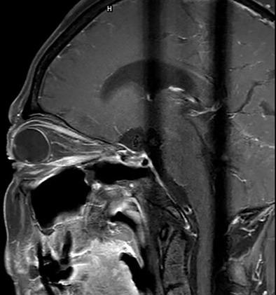
Latent intracranial meningeal metastases (IMM) of lung cancer is difficult to determine, yet it is critical to do so given that it impacts the treatment agent. Studies on this disease are rare, thus necessitating further investigation. As a case study, we will explore the application of optic neuroimaging in IMM. A 62-year-old female patient was diagnosed with lung adenocarcinoma, which had progressed to osseous metastasis. During the course of chemotherapy, the patient had bilateral vision loss and paralysis of extraocular muscles. Ophthalmologists ruled out disease of the retina and suspected intracranial metastasis; however, brain-enhanced magnetic resonance angiography and magnetic resonance venography were normal. Given the patient's severe osteoarthropathy and poor physical condition, she refused to undergo a lumbar puncture examination. Optic neuro-ophthalmology imaging was ultimately used. Utilizing optical coherence tomography, we found that the basement membrane layer in the papilledema was protruding up towards the vitreous cavity. To assist in visualization, the optic nerve sheath was enhanced with optic magnetic resonance imaging. With these methods, the dural metastasis was identified, the treatment agent was changed for the patient, and she had a successful recovery. Thus, optic neuro-ophthalmology imaging should be recommended for patients who are in the latent course of dural metastasis, and it could also be used to evaluate therapeutic efficacy.
中文翻译:

视神经眼科成像在肺癌潜伏脑膜转移中的应用
肺癌的潜在颅内脑膜转移 (IMM) 难以确定,但鉴于它会影响治疗药物,因此确定这一点至关重要。对这种疾病的研究很少,因此需要进一步研究。作为案例研究,我们将探讨视神经成像在 IMM 中的应用。一名 62 岁的女性患者被诊断为肺腺癌,已进展为骨转移。在化疗过程中,患者出现双侧视力下降和眼外肌麻痹。眼科医生排除了视网膜疾病和疑似颅内转移;然而,脑增强磁共振血管造影和磁共振静脉造影正常。鉴于患者骨关节病严重,身体状况不佳,拒绝接受腰穿检查。最终使用了光学神经眼科成像。利用光学相干断层扫描,我们发现视乳头水肿中的基底膜层向玻璃体腔突出。为了帮助可视化,用光学磁共振成像增强了视神经鞘。通过这些方法,确定了硬脑膜转移,为患者更换了治疗药物,她成功康复。因此,对于处于硬脑膜转移潜伏期的患者,应推荐视神经眼科成像,它也可用于评估治疗效果。为了帮助可视化,用光学磁共振成像增强了视神经鞘。通过这些方法,确定了硬脑膜转移,为患者更换了治疗药物,她成功康复。因此,对于处于硬脑膜转移潜伏期的患者,应推荐视神经眼科成像,它也可用于评估治疗效果。为了帮助可视化,用光学磁共振成像增强了视神经鞘。通过这些方法,确定了硬脑膜转移,为患者更换了治疗药物,她成功康复。因此,对于处于硬脑膜转移潜伏期的患者,应推荐视神经眼科成像,它也可用于评估治疗效果。
更新日期:2021-10-04
中文翻译:
视神经眼科成像在肺癌潜伏脑膜转移中的应用
肺癌的潜在颅内脑膜转移 (IMM) 难以确定,但鉴于它会影响治疗药物,因此确定这一点至关重要。对这种疾病的研究很少,因此需要进一步研究。作为案例研究,我们将探讨视神经成像在 IMM 中的应用。一名 62 岁的女性患者被诊断为肺腺癌,已进展为骨转移。在化疗过程中,患者出现双侧视力下降和眼外肌麻痹。眼科医生排除了视网膜疾病和疑似颅内转移;然而,脑增强磁共振血管造影和磁共振静脉造影正常。鉴于患者骨关节病严重,身体状况不佳,拒绝接受腰穿检查。最终使用了光学神经眼科成像。利用光学相干断层扫描,我们发现视乳头水肿中的基底膜层向玻璃体腔突出。为了帮助可视化,用光学磁共振成像增强了视神经鞘。通过这些方法,确定了硬脑膜转移,为患者更换了治疗药物,她成功康复。因此,对于处于硬脑膜转移潜伏期的患者,应推荐视神经眼科成像,它也可用于评估治疗效果。为了帮助可视化,用光学磁共振成像增强了视神经鞘。通过这些方法,确定了硬脑膜转移,为患者更换了治疗药物,她成功康复。因此,对于处于硬脑膜转移潜伏期的患者,应推荐视神经眼科成像,它也可用于评估治疗效果。为了帮助可视化,用光学磁共振成像增强了视神经鞘。通过这些方法,确定了硬脑膜转移,为患者更换了治疗药物,她成功康复。因此,对于处于硬脑膜转移潜伏期的患者,应推荐视神经眼科成像,它也可用于评估治疗效果。


 京公网安备 11010802027423号