当前位置:
X-MOL 学术
›
Mol. Pharmacol.
›
论文详情
Our official English website, www.x-mol.net, welcomes your
feedback! (Note: you will need to create a separate account there.)
High-Content Single-Cell Förster Resonance Energy Transfer Imaging of Cultured Striatal Neurons Reveals Novel Cross-Talk in the Regulation of Nuclear Signaling by Protein Kinase A and Extracellular Signal-Regulated Kinase 1/2
Molecular Pharmacology ( IF 3.2 ) Pub Date : 2021-12-01 , DOI: 10.1124/molpharm.121.000290 Jace Jones-Tabah 1 , Ryan D Martin 1 , Jason C Tanny 1 , Paul B S Clarke 2 , Terence E Hébert 2
Molecular Pharmacology ( IF 3.2 ) Pub Date : 2021-12-01 , DOI: 10.1124/molpharm.121.000290 Jace Jones-Tabah 1 , Ryan D Martin 1 , Jason C Tanny 1 , Paul B S Clarke 2 , Terence E Hébert 2
Affiliation
|
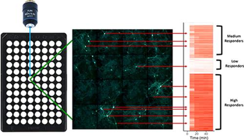
Genetically encoded biosensors can be used to track signaling events in living cells by measuring changes in fluorescence emitted by one or more fluorescent proteins. Here, we describe the use of genetically encoded biosensors based on Förster resonance energy transfer (FRET), combined with high-content microscopy, to image dynamic signaling events simultaneously in thousands of neurons in response to drug treatments. We first applied this approach to examine intercellular variation in signaling responses among cultured striatal neurons stimulated with multiple drugs. Using high-content FRET imaging and immunofluorescence, we identified neuronal subpopulations with unique responses to pharmacological manipulation and used nuclear morphology to identify medium spiny neurons within these heterogeneous striatal cultures. Focusing on protein kinase A (PKA) and extracellular signal-regulated kinase 1/2 (ERK1/2) signaling in the cytoplasm and nucleus, we noted pronounced intercellular differences among putative medium spiny neurons, in both the magnitude and kinetics of signaling responses to drug application. Importantly, a conventional “bulk” analysis that pooled all cells in culture yielded a different rank order of drug potency than that revealed by single-cell analysis. Using a single-cell analytical approach, we dissected the relative contributions of PKA and ERK1/2 signaling in striatal neurons and unexpectedly identified a novel role for ERK1/2 in promoting nuclear activation of PKA in striatal neurons. This finding adds a new dimension of signaling crosstalk between PKA and ERK1/2 with relevance to dopamine D1 receptor signaling in striatal neurons. In conclusion, high-content single-cell imaging can complement and extend traditional population-level analyses and provides a novel vantage point from which to study cellular signaling.
中文翻译:

培养的纹状体神经元的高内涵单细胞 Förster 共振能量转移成像揭示了蛋白激酶 A 和细胞外信号调节激酶 1/2 调节核信号的新串扰
遗传编码的生物传感器可用于通过测量一种或多种荧光蛋白发出的荧光变化来跟踪活细胞中的信号事件。在这里,我们描述了使用基于 Förster 共振能量转移 (FRET) 的基因编码生物传感器,结合高内涵显微镜,在数千个神经元中同时对药物治疗做出反应的动态信号事件成像。我们首先应用这种方法来检查用多种药物刺激的培养纹状体神经元之间信号反应的细胞间变化。使用高含量 FRET 成像和免疫荧光,我们确定了对药理学操作具有独特反应的神经元亚群,并使用核形态来确定这些异质纹状体培养物中的中等多刺神经元。关注细胞质和细胞核中的蛋白激酶 A (PKA) 和细胞外信号调节激酶 1/2 (ERK1/2) 信号,我们注意到推定的中等多刺神经元之间在信号响应的幅度和动力学方面存在显着的细胞间差异。药物应用。重要的是,将培养中的所有细胞汇集在一起的传统“批量”分析产生了与单细胞分析显示的药物效力不同的等级顺序。使用单细胞分析方法,我们剖析了纹状体神经元中 PKA 和 ERK1/2 信号传导的相对贡献,并意外地发现了 ERK1/2 在促进纹状体神经元中 PKA 核激活方面的新作用。这一发现增加了 PKA 和 ERK1/2 之间信号串扰的新维度,与纹状体神经元中的多巴胺 D1 受体信号传导相关。
更新日期:2021-11-20
中文翻译:
培养的纹状体神经元的高内涵单细胞 Förster 共振能量转移成像揭示了蛋白激酶 A 和细胞外信号调节激酶 1/2 调节核信号的新串扰
遗传编码的生物传感器可用于通过测量一种或多种荧光蛋白发出的荧光变化来跟踪活细胞中的信号事件。在这里,我们描述了使用基于 Förster 共振能量转移 (FRET) 的基因编码生物传感器,结合高内涵显微镜,在数千个神经元中同时对药物治疗做出反应的动态信号事件成像。我们首先应用这种方法来检查用多种药物刺激的培养纹状体神经元之间信号反应的细胞间变化。使用高含量 FRET 成像和免疫荧光,我们确定了对药理学操作具有独特反应的神经元亚群,并使用核形态来确定这些异质纹状体培养物中的中等多刺神经元。关注细胞质和细胞核中的蛋白激酶 A (PKA) 和细胞外信号调节激酶 1/2 (ERK1/2) 信号,我们注意到推定的中等多刺神经元之间在信号响应的幅度和动力学方面存在显着的细胞间差异。药物应用。重要的是,将培养中的所有细胞汇集在一起的传统“批量”分析产生了与单细胞分析显示的药物效力不同的等级顺序。使用单细胞分析方法,我们剖析了纹状体神经元中 PKA 和 ERK1/2 信号传导的相对贡献,并意外地发现了 ERK1/2 在促进纹状体神经元中 PKA 核激活方面的新作用。这一发现增加了 PKA 和 ERK1/2 之间信号串扰的新维度,与纹状体神经元中的多巴胺 D1 受体信号传导相关。


 京公网安备 11010802027423号