Our official English website, www.x-mol.net, welcomes your
feedback! (Note: you will need to create a separate account there.)
Micro-Raman spectroscopy of lipid halo and dense-core amyloid plaques: aging process characterization in the Alzheimer's disease APPswePS1ΔE9 mouse model
Analyst ( IF 3.6 ) Pub Date : 2021-08-11 , DOI: 10.1039/d1an01078f Emerson A Fonseca 1, 2 , Lucas Lafeta 1 , João Luiz Campos 1 , Renan Cunha 1 , Alexandre Barbosa 1, 3 , Marco A Romano-Silva 4 , Rafael Vieira 5 , Leandro M Malard 1 , Ado Jorio 1, 2
Analyst ( IF 3.6 ) Pub Date : 2021-08-11 , DOI: 10.1039/d1an01078f Emerson A Fonseca 1, 2 , Lucas Lafeta 1 , João Luiz Campos 1 , Renan Cunha 1 , Alexandre Barbosa 1, 3 , Marco A Romano-Silva 4 , Rafael Vieira 5 , Leandro M Malard 1 , Ado Jorio 1, 2
Affiliation
|
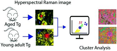
The deposition of amyloid plaques is considered one of the main microscopic features of Alzheimer's disease (AD). Since plaque formation can precede extensive neurodegeneration and it is the main clinical manifestation of AD, it constitutes a relevant target for new treatment and diagnostic approaches. Micro-Raman spectroscopy, a label-free technique, is an accurate method for amyloid plaque identification and characterization. Here, we present a high spatial resolution micro-Raman hyperspectral study in transgenic APPswePS1ΔE9 mouse brains, showing details of AD tissue biochemical and histological changes without staining. First we used stimulated micro-Raman scattering to identify the lipid-rich halo surrounding the amyloid plaque, and then proceeded with spontaneous (conventional) micro-Raman spectral mapping, which shows a cholesterol and sphingomyelin lipid-rich halo structure around dense-core amyloid plaques. The detailed images of this lipid halo relate morphologically well with dystrophic neurites surrounding plaques. Principal Component Analysis (PCA) of the micro-Raman hyperspectral data indicates the feasibility of the optical biomarkers of AD progression with the potential for discriminating transgenic groups of young adult mice (6-month-old) from older ones (12-month-old). Frequency-specific PCA suggests that plaque-related neurodegeneration is the predominant change captured by Raman spectroscopy, and the main differences are highlighted by vibrational modes associated with cholesterol located majorly in the lipid halo.
中文翻译:

脂质晕和致密核心淀粉样蛋白斑块的微拉曼光谱:阿尔茨海默病 APPswePS1ΔE9 小鼠模型中的衰老过程表征
淀粉样斑块的沉积被认为是阿尔茨海默病 (AD) 的主要微观特征之一。由于斑块形成可以先于广泛的神经变性并且是 AD 的主要临床表现,因此它构成了新治疗和诊断方法的相关目标。微拉曼光谱是一种无标记技术,是一种准确的淀粉样斑块识别和表征方法。在这里,我们在转基因 APPswePS1ΔE9 小鼠大脑中进行了高空间分辨率微拉曼高光谱研究,显示了 AD 组织生化和组织学变化的细节,无需染色。首先,我们使用受激微拉曼散射来识别淀粉样斑块周围的富含脂质的晕圈,然后进行自发(常规)微拉曼光谱映射,它显示了密集核心淀粉样斑块周围的胆固醇和鞘磷脂富含脂质的光环结构。这种脂质晕的详细图像在形态学上与斑块周围的营养不良性神经突相关。微拉曼高光谱数据的主成分分析 (PCA) 表明 AD 进展的光学生物标志物的可行性,具有区分年轻成年小鼠(6 个月大)和年长小鼠(12 个月大)转基因组的潜力)。频率特异性 PCA 表明斑块相关的神经变性是拉曼光谱捕获的主要变化,主要差异通过与主要位于脂质晕中的胆固醇相关的振动模式突出显示。这种脂质晕的详细图像在形态学上与斑块周围的营养不良性神经突相关。微拉曼高光谱数据的主成分分析 (PCA) 表明 AD 进展的光学生物标志物的可行性,具有区分年轻成年小鼠(6 个月大)和年长小鼠(12 个月大)转基因组的潜力)。频率特异性 PCA 表明斑块相关的神经变性是拉曼光谱捕获的主要变化,主要差异通过与主要位于脂质晕中的胆固醇相关的振动模式突出显示。这种脂质晕的详细图像在形态学上与斑块周围的营养不良性神经突相关。微拉曼高光谱数据的主成分分析 (PCA) 表明 AD 进展的光学生物标志物的可行性,具有区分年轻成年小鼠(6 个月大)和年长小鼠(12 个月大)转基因组的潜力)。频率特异性 PCA 表明斑块相关的神经变性是拉曼光谱捕获的主要变化,主要差异通过与主要位于脂质晕中的胆固醇相关的振动模式突出显示。微拉曼高光谱数据的主成分分析 (PCA) 表明 AD 进展的光学生物标志物的可行性,具有区分年轻成年小鼠(6 个月大)和年长小鼠(12 个月大)转基因组的潜力)。频率特异性 PCA 表明斑块相关的神经变性是拉曼光谱捕获的主要变化,主要差异通过与主要位于脂质晕中的胆固醇相关的振动模式突出显示。微拉曼高光谱数据的主成分分析 (PCA) 表明 AD 进展的光学生物标志物的可行性,具有区分年轻成年小鼠(6 个月大)和年长小鼠(12 个月大)转基因组的潜力)。频率特异性 PCA 表明斑块相关的神经变性是拉曼光谱捕获的主要变化,主要差异通过与主要位于脂质晕中的胆固醇相关的振动模式突出显示。
更新日期:2021-09-10
中文翻译:
脂质晕和致密核心淀粉样蛋白斑块的微拉曼光谱:阿尔茨海默病 APPswePS1ΔE9 小鼠模型中的衰老过程表征
淀粉样斑块的沉积被认为是阿尔茨海默病 (AD) 的主要微观特征之一。由于斑块形成可以先于广泛的神经变性并且是 AD 的主要临床表现,因此它构成了新治疗和诊断方法的相关目标。微拉曼光谱是一种无标记技术,是一种准确的淀粉样斑块识别和表征方法。在这里,我们在转基因 APPswePS1ΔE9 小鼠大脑中进行了高空间分辨率微拉曼高光谱研究,显示了 AD 组织生化和组织学变化的细节,无需染色。首先,我们使用受激微拉曼散射来识别淀粉样斑块周围的富含脂质的晕圈,然后进行自发(常规)微拉曼光谱映射,它显示了密集核心淀粉样斑块周围的胆固醇和鞘磷脂富含脂质的光环结构。这种脂质晕的详细图像在形态学上与斑块周围的营养不良性神经突相关。微拉曼高光谱数据的主成分分析 (PCA) 表明 AD 进展的光学生物标志物的可行性,具有区分年轻成年小鼠(6 个月大)和年长小鼠(12 个月大)转基因组的潜力)。频率特异性 PCA 表明斑块相关的神经变性是拉曼光谱捕获的主要变化,主要差异通过与主要位于脂质晕中的胆固醇相关的振动模式突出显示。这种脂质晕的详细图像在形态学上与斑块周围的营养不良性神经突相关。微拉曼高光谱数据的主成分分析 (PCA) 表明 AD 进展的光学生物标志物的可行性,具有区分年轻成年小鼠(6 个月大)和年长小鼠(12 个月大)转基因组的潜力)。频率特异性 PCA 表明斑块相关的神经变性是拉曼光谱捕获的主要变化,主要差异通过与主要位于脂质晕中的胆固醇相关的振动模式突出显示。这种脂质晕的详细图像在形态学上与斑块周围的营养不良性神经突相关。微拉曼高光谱数据的主成分分析 (PCA) 表明 AD 进展的光学生物标志物的可行性,具有区分年轻成年小鼠(6 个月大)和年长小鼠(12 个月大)转基因组的潜力)。频率特异性 PCA 表明斑块相关的神经变性是拉曼光谱捕获的主要变化,主要差异通过与主要位于脂质晕中的胆固醇相关的振动模式突出显示。微拉曼高光谱数据的主成分分析 (PCA) 表明 AD 进展的光学生物标志物的可行性,具有区分年轻成年小鼠(6 个月大)和年长小鼠(12 个月大)转基因组的潜力)。频率特异性 PCA 表明斑块相关的神经变性是拉曼光谱捕获的主要变化,主要差异通过与主要位于脂质晕中的胆固醇相关的振动模式突出显示。微拉曼高光谱数据的主成分分析 (PCA) 表明 AD 进展的光学生物标志物的可行性,具有区分年轻成年小鼠(6 个月大)和年长小鼠(12 个月大)转基因组的潜力)。频率特异性 PCA 表明斑块相关的神经变性是拉曼光谱捕获的主要变化,主要差异通过与主要位于脂质晕中的胆固醇相关的振动模式突出显示。


 京公网安备 11010802027423号