Journal of Microbiological Methods ( IF 1.7 ) Pub Date : 2021-09-10 , DOI: 10.1016/j.mimet.2021.106327 Shinsuke Nerome 1 , Naoki Yokota 1 , Yoshihiro Ojima 1 , Masayuki Azuma 1
|
Introduction
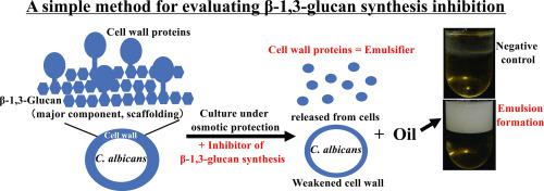
The cell wall β-1,3-glucan of fungal pathogen Candida albicans is an attractive antifungal target. β-1,3-Glucan is the skeletal structure in the cell wall and the major scaffold for cell wall proteins. In previous studies using Saccharomyces cerevisiae, strong emulsification was detected by mixing cell wall proteins with oil. To date, there have been no reports of applying an emulsification phenomenon to assessing β-1,3-glucan synthesis inhibition.
Objective
The aim of this study was to clarify that emulsification is useful as an indicator for evaluating β-1,3-glucan synthesis inhibition in C. albicans.
Methods
At first, whether cell wall proteins released from cells by β-1,3-glucanase treatment worked as an effective emulsifier in C. albicans was examined. Next, whether emulsification occurred even in the culture supernatant brought about by treating with bioactive compounds, including β-1,3-glucan synthesis inhibitors, under osmotic protection was investigated. In addition, the release of cell wall proteins into the culture medium by treating with those compounds was examined. Finally, a simpler evaluation method using emulsion formation was examined for application to screening of inhibitors.
Results
Emulsification occurred by cell wall proteins obtained by treating with β-1,3-glucanase in C. albicans. In addition, cell wall proteins were released into the culture medium by treating with β-1,3-glucan synthesis inhibitors, resulting in emulsification. However, such phenomena were not observed in the case of other bioactive compounds. Furthermore, emulsification could be detected in the culture broth obtained by static culture on a small scale.
Conclusions
The obtained results strongly implied that emulsification results from decreased β-1,3-glucan levels in the cell wall. As emulsification can be simply evaluated by mixing the culture broth with oil, in the future application to the initial assessment and screening of β-1,3-glucan synthesis inhibitors is expected.
中文翻译:
以乳状液形成为指标评价 β-1,3-葡聚糖合成抑制
介绍
真菌病原体白色念珠菌的细胞壁β-1,3-葡聚糖是一种有吸引力的抗真菌靶标。β-1,3-葡聚糖是细胞壁中的骨架结构和细胞壁蛋白的主要支架。在以前使用酿酒酵母的研究中,通过将细胞壁蛋白与油混合来检测到强乳化作用。迄今为止,还没有关于应用乳化现象来评估 β-1,3-葡聚糖合成抑制的报道。
客观的
本研究的目的是阐明乳化可用作评估白色念珠菌中 β-1,3-葡聚糖合成抑制的指标。
方法
首先,检查了通过 β-1,3-葡聚糖酶处理从细胞中释放的细胞壁蛋白是否在白色念珠菌中作为有效的乳化剂起作用。接下来,研究了在渗透保护下是否甚至在用生物活性化合物(包括β-1,3-葡聚糖合成抑制剂)处理所产生的培养上清液中发生乳化。此外,检查了通过用这些化合物处理细胞壁蛋白释放到培养基中的情况。最后,研究了一种使用乳液形成的更简单的评估方法,用于筛选抑制剂。
结果
通过用β-1,3-葡聚糖酶处理白色念珠菌获得的细胞壁蛋白发生乳化。此外,通过用β-1,3-葡聚糖合成抑制剂处理,细胞壁蛋白释放到培养基中,导致乳化。然而,在其他生物活性化合物的情况下没有观察到这种现象。此外,可以在小规模静态培养获得的培养液中检测到乳化。
结论
所得结果强烈暗示乳化是由于细胞壁中β-1,3-葡聚糖水平降低所致。由于可以通过将培养液与油混合来简单地评估乳化作用,因此预计未来将应用于 β-1,3-葡聚糖合成抑制剂的初步评估和筛选。


 京公网安备 11010802027423号