Surfaces and Interfaces ( IF 5.7 ) Pub Date : 2021-09-04 , DOI: 10.1016/j.surfin.2021.101439 R.T. Konatu 1 , D.D. Domingues 2 , A.L.A. Escada 1 , J.A.M. Chaves 3 , M.F.D. Netipanyj 4 , R.Z. Nakazato 1 , K.C. Popat 5 , C.R. Grandini 6 , A.P.R. Alves Claro 1, 2
|
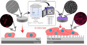
In recent years, studies have been shown that the presence of TiO2 nanotubes on the titanium alloy surfaces could induce the enhancement of the cell adhesion on the titanium alloys surface. In the present study, the cell response of the surface of the Ti-15Zr alloy after TiO2 nanotubes growth (NTs) via anodic oxidation was evaluated. TiO2 NTs were obtained in organic electrolytes and analyzed after annealing using Scanning electron microscopy (SEM), X-ray diffraction (XRD), and Raman spectroscopy. The attachment and growth of adult human adipose-derived stem cells (ADSCs) were considered for 1 and 7 days for better annealing conditions (450°C). Besides, the influence of the TiO2 NTs growth on the corrosion resistance and Staphylococcus epidermidis bacterial adhesion was evaluated. Results indicated that a self-organized and homogeneous layer of TiO2 nanotubes was formed. XRD analysis and SEM micrographs confirmed that as-anodized, amorphous nanotubes crystallized into anatase phase at 450°C. According to electrochemical analysis, Ti-15Zr alloy exhibited better corrosion resistance compared to commercially pure titanium (cp Ti), but there was no significant difference in passivation capacity between samples evaluated before and after anodization. It was observed that an increase in cellular adhesion and no significant difference in bacterial proliferation occurred despite the presence of TiO2 nanotubes which changed the surface roughness.
中文翻译:
在 Ti-15Zr 合金表面生长自组织 TiO2 纳米管以增强细胞响应的合成和表征
近年来的研究表明,钛合金表面TiO 2纳米管的存在可以增强钛合金表面的细胞粘附。在本研究中,评估了通过阳极氧化生长TiO 2纳米管 (NTs)后 Ti-15Zr 合金表面的电池响应。TiO 2 NTs 在有机电解质中获得,并在退火后使用扫描电子显微镜 (SEM)、X 射线衍射 (XRD) 和拉曼光谱进行分析。成人脂肪干细胞 (ADSC) 的附着和生长被考虑为 1 天和 7 天,以获得更好的退火条件 (450°C)。此外,TiO 2 NTs 的生长对耐蚀性和评价表皮葡萄球菌细菌粘附。结果表明,形成了自组织且均质的TiO 2纳米管层。XRD 分析和 SEM 显微照片证实,阳极氧化的无定形纳米管在 450°C 下结晶成锐钛矿相。根据电化学分析,与商业纯钛 (cp Ti) 相比,Ti-15Zr 合金表现出更好的耐腐蚀性,但阳极氧化前后评估的样品之间的钝化能力没有显着差异。据观察,尽管存在改变表面粗糙度的 TiO 2纳米管,但细胞粘附增加并且细菌增殖没有显着差异。


 京公网安备 11010802027423号