当前位置:
X-MOL 学术
›
Ann. N. Y. Acad. Sci.
›
论文详情
Our official English website, www.x-mol.net, welcomes your
feedback! (Note: you will need to create a separate account there.)
Use of Mayer wave activity to demonstrate aberrant cardiovascular autonomic control following sports concussion injury
Annals of the New York Academy of Sciences ( IF 4.1 ) Pub Date : 2021-09-03 , DOI: 10.1111/nyas.14683 Michael F La Fountaine 1, 2, 3 , Asante N Hohn 1, 4 , Caroline L Leahy 1, 5 , Anthony J Testa 6 , Joseph P Weir 7, 8
Annals of the New York Academy of Sciences ( IF 4.1 ) Pub Date : 2021-09-03 , DOI: 10.1111/nyas.14683 Michael F La Fountaine 1, 2, 3 , Asante N Hohn 1, 4 , Caroline L Leahy 1, 5 , Anthony J Testa 6 , Joseph P Weir 7, 8
Affiliation
|
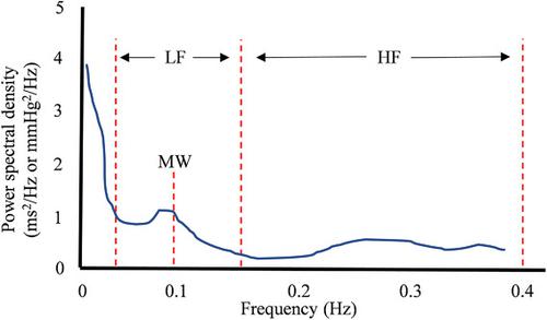
Dysregulation of cardiovascular autonomic control is gaining recognition as a prevailing consequence of concussion injury. Characterizing the presence of autonomic dysfunction in concussed persons is inconsistent and conventional metrics of autonomic function cannot differentiate the presence/absence of injury. Mayer wave (MW) activity originates through baroreflex adjustments to blood pressure (BP) oscillations that appear in the low-frequency (LF: 0.04–0.15 Hz) band of the BP and heart rate (HR) power spectrum after a fast Fourier transform. We prospectively explored MW activity (∼0.1 Hz) in 19 concussed and 19 noninjured athletes for 5 min while seated at rest within 48 h and 1 week of injury. MW activity was derived from the LF band of continuous digital electrocardiogram and beat-to-beat BP signals (LFHR, LF-SBP, MWHR, and MW-SBP, respectively); a proportion between MWBP and MWHR was computed (cMW). At 48 h, the concussion group had a significantly lower MWBP and cMW than controls; these differences were gone by 1 week. MWHR, LFHR, and LF-SBP were not different between groups at either visit. Attenuated sympathetic vasomotor tone was present and the central autonomic mechanisms regulating MW activity to the heart and peripheral vasculature became transiently discordant early after concussion with apparent resolution by 1 week.
中文翻译:

使用 Mayer 波活动来证明运动性脑震荡损伤后的异常心血管自主控制
心血管自主神经控制失调作为脑震荡损伤的普遍后果得到了认可。描述脑震荡者存在自主神经功能障碍的特征是不一致的,自主神经功能的常规指标无法区分损伤的存在/不存在。迈耶波 (MW) 活动源自对血压 (BP) 振荡的压力反射调整,该振荡出现在快速傅立叶变换后 BP 和心率 (HR) 功率谱的低频 (LF: 0.04–0.15 Hz) 频带中。我们前瞻性地研究了 19 名脑震荡和 19 名未受伤运动员的 MW 活动(~0.1 Hz),持续 5 分钟,同时在受伤后 48 小时和 1 周内静坐。MW 活动来自连续数字心电图的 LF 波段和逐搏 BP 信号(LFHR、LF-SBP、MWHR 和 MW-SBP,分别); 计算了 MWBP 和 MWHR 之间的比例 (cMW)。48 h时,脑震荡组MWBP和cMW明显低于对照组;这些差异在 1 周内消失了。MWHR、LFHR 和 LF-SBP 在任何一次访问中各组之间均无差异。存在减弱的交感神经血管舒缩张力,调节 MW 对心脏和外周脉管系统的活动的中枢自主神经机制在脑震荡后早期变得短暂不协调,并在 1 周时明显消退。
更新日期:2021-09-03
中文翻译:
使用 Mayer 波活动来证明运动性脑震荡损伤后的异常心血管自主控制
心血管自主神经控制失调作为脑震荡损伤的普遍后果得到了认可。描述脑震荡者存在自主神经功能障碍的特征是不一致的,自主神经功能的常规指标无法区分损伤的存在/不存在。迈耶波 (MW) 活动源自对血压 (BP) 振荡的压力反射调整,该振荡出现在快速傅立叶变换后 BP 和心率 (HR) 功率谱的低频 (LF: 0.04–0.15 Hz) 频带中。我们前瞻性地研究了 19 名脑震荡和 19 名未受伤运动员的 MW 活动(~0.1 Hz),持续 5 分钟,同时在受伤后 48 小时和 1 周内静坐。MW 活动来自连续数字心电图的 LF 波段和逐搏 BP 信号(LFHR、LF-SBP、MWHR 和 MW-SBP,分别); 计算了 MWBP 和 MWHR 之间的比例 (cMW)。48 h时,脑震荡组MWBP和cMW明显低于对照组;这些差异在 1 周内消失了。MWHR、LFHR 和 LF-SBP 在任何一次访问中各组之间均无差异。存在减弱的交感神经血管舒缩张力,调节 MW 对心脏和外周脉管系统的活动的中枢自主神经机制在脑震荡后早期变得短暂不协调,并在 1 周时明显消退。


 京公网安备 11010802027423号