NeuroImage: Clinical ( IF 3.4 ) Pub Date : 2021-09-02 , DOI: 10.1016/j.nicl.2021.102817 Veronica Ravano 1 , Michaela Andelova 2 , Mário João Fartaria 1 , Mazen Fouad A-Wali Mahdi 3 , Bénédicte Maréchal 1 , Reto Meuli 4 , Tomas Uher 2 , Jan Krasensky 5 , Manuela Vaneckova 5 , Dana Horakova 2 , Tobias Kober 1 , Jonas Richiardi 4
|
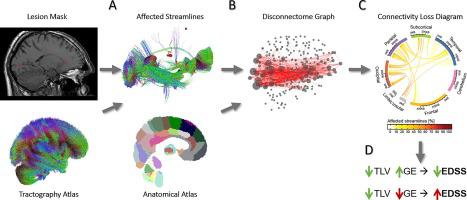
The translational potential of MR-based connectivity modelling is limited by the need for advanced diffusion imaging, which is not part of clinical protocols for many diseases. In addition, where diffusion data is available, brain connectivity analyses rely on tractography algorithms which imply two major limitations. First, tracking algorithms are known to be sensitive to the presence of white matter lesions and therefore leading to interpretation pitfalls and poor inter-subject comparability in clinical applications such as multiple sclerosis. Second, tractography quality is highly dependent on the acquisition parameters of diffusion sequences, leading to a trade-off between acquisition time and tractography precision.
Here, we propose an atlas-based approach to study the interplay between structural disconnectivity and lesions without requiring individual diffusion imaging. In a multi-centric setting involving three distinct multiple sclerosis datasets (containing both 1.5 T and 3 T data), we compare our atlas-based structural disconnectome computation pipeline to disconnectomes extracted from individual tractography and explore its clinical utility for reducing the gap between radiological findings and clinical symptoms in multiple sclerosis. Results using topological graph properties showed that overall, our atlas-based disconnectomes were suitable approximations of individual disconnectomes from diffusion imaging. Small-worldness was found to decrease for larger total lesion volumes thereby suggesting a loss of efficiency in brain connectivity of MS patients. Finally, the global efficiency of the created brain graph, combined with total lesion volume, allowed to stratify patients into subgroups with different clinical scores in all three cohorts.
中文翻译:
验证多发性硬化症中基于图谱的病变断开组学:一项回顾性多中心研究
基于 MR 的连通性建模的转化潜力受到对高级扩散成像的需求的限制,这不是许多疾病临床协议的一部分。此外,在可获得扩散数据的情况下,大脑连接分析依赖于牵张图算法,这意味着两个主要限制。首先,众所周知,跟踪算法对白质病变的存在很敏感,因此在多发性硬化症等临床应用中会导致解释缺陷和主体间可比性差。其次,纤维束成像质量高度依赖于扩散序列的采集参数,导致采集时间和纤维束成像精度之间的权衡。
在这里,我们提出了一种基于图谱的方法来研究结构断开性和病变之间的相互作用,而不需要单独的扩散成像。在涉及三个不同的多发性硬化症数据集(包含 1.5 T 和 3 T 数据)的多中心环境中,我们将基于图谱的结构断开组计算管道与从单个纤维束成像中提取的断开组进行比较,并探索其在减少放射学之间差距的临床效用多发性硬化症的发现和临床症状。使用拓扑图属性的结果表明,总体而言,我们基于图谱的断开组是扩散成像中单个断开组的合适近似值。发现总病变体积较大时小世界性会降低,从而表明 MS 患者的大脑连接效率降低。


 京公网安备 11010802027423号