Our official English website, www.x-mol.net, welcomes your
feedback! (Note: you will need to create a separate account there.)
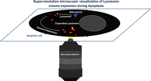
Super-Resolution Microscopy Revealed the Lysosomal Expansion During Epigallocatechin Gallate-Mediated Apoptosis
Langmuir ( IF 3.7 ) Pub Date : 2021-09-01 , DOI: 10.1021/acs.langmuir.1c01742 Pushpendra M Mishra 1, 2, 3 , Chethana Rao 1, 2 , Ankita Sarkar 1, 3 , Aditya Yadav 1, 2 , Kush Kaushik 1, 2 , Amit Jaiswal 1, 3 , Chayan K Nandi 1, 2, 3
Langmuir ( IF 3.7 ) Pub Date : 2021-09-01 , DOI: 10.1021/acs.langmuir.1c01742 Pushpendra M Mishra 1, 2, 3 , Chethana Rao 1, 2 , Ankita Sarkar 1, 3 , Aditya Yadav 1, 2 , Kush Kaushik 1, 2 , Amit Jaiswal 1, 3 , Chayan K Nandi 1, 2, 3
Affiliation
|
Direct visualization of the dynamic events in lysosomes during drug-mediated programmed cell death (apoptosis) is a great challenge. This is due to the lack of resolving power of a conventional microscope and also the unavailability of a suitable multimodal probe that simultaneously can carry the drug with high loading capacity and ensure its specific internalization into lysosomes. In this work, using super-resolution microscopy, we observed the lysosomal expansion during apoptosis that was treated with epigallocatechin gallate (EGCG) conjugated to bovine serum albumin (BSA). Albumin protein is known to internalize into lysosomes via endocytosis, thus helping in the specific delivery of EGCG to the lysosomal compartment. The conjugation of EGCG to BSA not only helped in increasing the killing efficiency of cancer cells but it also reduces the side effects and produces minimal reactive oxygen species. The decrease in local viscosity helped in lysosomal expansion during apoptosis.
中文翻译:

超分辨率显微镜揭示了表没食子儿茶素没食子酸酯介导的细胞凋亡过程中溶酶体的扩张
在药物介导的程序性细胞死亡(细胞凋亡)期间溶酶体中动态事件的直接可视化是一个巨大的挑战。这是由于缺乏传统显微镜的分辨能力,也没有合适的多模态探针可以同时携带具有高负载能力的药物并确保其特异性内化到溶酶体中。在这项工作中,我们使用超分辨率显微镜观察了细胞凋亡过程中溶酶体的扩张,该细胞用与牛血清白蛋白 (BSA) 结合的表没食子儿茶素没食子酸酯 (EGCG) 处理。已知白蛋白蛋白通过内吞作用内化到溶酶体中,从而有助于将 EGCG 特异性递送到溶酶体区室。EGCG 与 BSA 的结合不仅有助于提高癌细胞的杀伤效率,而且还减少了副作用并产生最少的活性氧。局部粘度的降低有助于细胞凋亡过程中溶酶体的扩张。
更新日期:2021-09-14
中文翻译:
超分辨率显微镜揭示了表没食子儿茶素没食子酸酯介导的细胞凋亡过程中溶酶体的扩张
在药物介导的程序性细胞死亡(细胞凋亡)期间溶酶体中动态事件的直接可视化是一个巨大的挑战。这是由于缺乏传统显微镜的分辨能力,也没有合适的多模态探针可以同时携带具有高负载能力的药物并确保其特异性内化到溶酶体中。在这项工作中,我们使用超分辨率显微镜观察了细胞凋亡过程中溶酶体的扩张,该细胞用与牛血清白蛋白 (BSA) 结合的表没食子儿茶素没食子酸酯 (EGCG) 处理。已知白蛋白蛋白通过内吞作用内化到溶酶体中,从而有助于将 EGCG 特异性递送到溶酶体区室。EGCG 与 BSA 的结合不仅有助于提高癌细胞的杀伤效率,而且还减少了副作用并产生最少的活性氧。局部粘度的降低有助于细胞凋亡过程中溶酶体的扩张。


 京公网安备 11010802027423号