Colloids and Surfaces B: Biointerfaces ( IF 5.4 ) Pub Date : 2021-09-01 , DOI: 10.1016/j.colsurfb.2021.112094 Angshuman Bharadwaz 1 , Ambalangodage C Jayasuriya 2
|
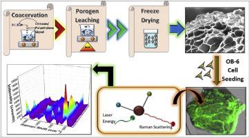
Porous chitosan (CS) particles were fabricated using a novel two-step technique that employed a porogen leaching phase followed by lyophilization or freeze-drying. Poly(ethylene glycol) (PEG) was mixed as a porogen in two different quantities with the CS solution before particle synthesis via coacervation. After the PEG leached out into deionized (DI) water at an elevated constant temperature, the final freeze-dried CS particles revealed surface features that resembled pore pockets. A three-dimensional (3D) culture of murine osteoblast cell line (OB-6) was seeded on these particles to analyze the effect of the porous structure on the cell activity, as compared to a control group with no added porogen. The results highlighted an enhancement in cell adhesion and proliferation on the two porous sample groups. A Raman spectroscopy-based label-free technique for live cell biomarker analysis was applied using multivariate spectral analysis. Results of the spectral analysis in the molecular fingerprint region corresponding to the Raman shift between 900 cm−1 and 1700 cm−1inferred inter-group variations. The bands at 1005 cm−1 and 1375 cm−1 were assigned to the live cell biomarkers phenylalanine and glycosaminoglycan, respectively, and were assessed during the multivariate spectral analysis. The corresponding score plot and loading information generated from the Principal Component Analysis (PCA) of the Raman spectrum at day 7 and day 14, pointed at inter-group spectral variations related to cell adhesion and proliferation between the two porous CS particle groups and the control.
中文翻译:
使用新型两步致孔剂浸出和冻干方法制备多孔壳聚糖颗粒,并对活粘附细胞进行无标记多变量光谱评估
多孔壳聚糖 (CS) 颗粒是使用一种新的两步技术制造的,该技术采用了致孔剂浸出阶段,然后是冻干或冷冻干燥。聚(乙二醇)(PEG)作为致孔剂以两种不同的量与 CS 溶液混合,然后通过凝聚进行颗粒合成。在 PEG 在升高的恒温下浸出到去离子 (DI) 水中后,最终冻干的 CS 颗粒显示出类似于孔袋的表面特征。与未添加致孔剂的对照组相比,将小鼠成骨细胞系 (OB-6) 的三维 (3D) 培养物接种在这些颗粒上,以分析多孔结构对细胞活性的影响。结果突出了两个多孔样品组的细胞粘附和增殖的增强。使用多变量光谱分析应用基于拉曼光谱的无标记技术进行活细胞生物标志物分析。900 cm 之间拉曼位移对应的分子指纹区光谱分析结果-1和 1700 cm -1推断组间变异。1005 cm -1和1375 cm -1处的条带分别分配给活细胞生物标志物苯丙氨酸和糖胺聚糖,并在多变量光谱分析期间进行评估。从第 7 天和第 14 天的拉曼光谱的主成分分析 (PCA) 生成的相应得分图和加载信息指出了与两个多孔 CS 颗粒组和对照之间的细胞粘附和增殖相关的组间光谱变化.


 京公网安备 11010802027423号