Clinical Research in Cardiology ( IF 3.8 ) Pub Date : 2021-08-26 , DOI: 10.1007/s00392-021-01929-5 Philipp Breitbart 1, 2, 3 , Alexander Koch 1, 3 , Marco Schmidt 1, 3 , Annett Magedanz 1, 3 , Edelgard Lindhoff-Last 1, 3 , Thomas Voigtländer 1, 3 , Axel Schmermund 1, 3 , Rajendra H Mehta 4 , Holger Eggebrecht 1, 3
|
Objectives
We assessed possible myocardial involvement in previously cardiac healthy post-COVID patients referred for persisting symptoms with suspected myocarditis.
Background
Prior studies suggested myocardial inflammation in patients with coronavirus-induced disease 2019 (COVID-19). However, the prevalence of cardiac involvement among COVID patients varied between 1.4 and 78%.
Methods
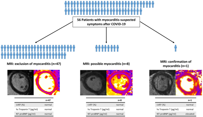
A total of 56 post-COVID patients without previous heart diseases were included consecutively into this study. All patients had positive antibody titers against SARS-CoV-2. Patients were referred for persistent symptoms such as chest pain/discomfort, shortness of breath, or intolerance to activity. All patients underwent standardized cardiac assessment including electrocardiogram (ECG), cardiac biomarkers, echocardiography, and cardiac magnetic resonance (CMR).
Results
56 Patients (46 ± 12 years, 54% females) presented 71 ± 66 days after their COVID-19 disease. In most patients, the course of COVID-19 was mild, with hospital treatment being necessary in five (9%). At presentation, patients most often reported persistent fatigue (75%), chest pain (71%), and shortness of breath (66%). Acute myocarditis was confirmed by T1/T2-weighed CMR and elevated NTpro-BNP levels in a single patient (2%). Left ventricular ejection fraction was 56% in this patient. Additional eight patients (14%) showed suspicious CMR findings, including myocardial edema without fibrosis (n = 3), or non-ischemic myocardial injury suggesting previous inflammation (n = 5). However, myocarditis could ultimately not be confirmed according to 2018 Lake Louise criteria; ECG, echo and lab findings were inconspicuous in all eight patients.
Conclusions
Among 56 post-COVID patients with persistent thoracic complaints final diagnosis of myocarditis could be confirmed in a single patient using CMR.
Graphic abstract
中文翻译:
COVID 后疑似心肌炎患者的临床和心脏磁共振检查结果
目标
我们评估了先前心脏健康的 COVID 后患者可能的心肌受累,这些患者因疑似心肌炎的持续症状而转诊。
背景
先前的研究表明,2019 年冠状病毒引起的疾病 (COVID-19) 患者存在心肌炎症。然而,COVID 患者中心脏受累的患病率在 1.4% 到 78% 之间变化。
方法
共有 56 名既往没有心脏病的 COVID 后患者被连续纳入本研究。所有患者的 SARS-CoV-2 抗体滴度均为阳性。患者因持续症状而被转诊,例如胸痛/不适、呼吸急促或对活动不耐受。所有患者均接受了标准化的心脏评估,包括心电图(ECG)、心脏生物标志物、超声心动图和心脏磁共振(CMR)。
结果
56 名患者(46 ± 12 岁,54% 女性)在感染 COVID-19 后 71 ± 66 天就诊。在大多数患者中,COVID-19 的病程较轻,有 5 人 (9%) 需要住院治疗。就诊时,患者最常报告持续疲劳(75%)、胸痛(71%)和呼吸急促(66%)。T1/T2 加权 CMR 和单个患者的 NTpro-BNP 水平升高证实了急性心肌炎 (2%)。该患者的左心室射血分数为 56%。另外 8 名患者 (14%) 表现出可疑的 CMR 发现,包括无纤维化的心肌水肿 ( n = 3),或提示先前炎症的非缺血性心肌损伤 ( n = 5)。但根据 2018 年路易斯湖标准最终无法确诊心肌炎;所有 8 名患者的心电图、回声和实验室检查结果均不明显。
结论
在 56 名 COVID 后持续胸部不适的患者中,可以使用 CMR 在单个患者中确认心肌炎的最终诊断。


 京公网安备 11010802027423号