Cell Stem Cell ( IF 19.8 ) Pub Date : 2021-08-17 , DOI: 10.1016/j.stem.2021.07.010 Elke Gabriel 1 , Walid Albanna 2 , Giovanni Pasquini 3 , Anand Ramani 1 , Natasa Josipovic 4 , Aruljothi Mariappan 1 , Friedrich Schinzel 1 , Celeste M Karch 5 , Guobin Bao 6 , Marco Gottardo 1 , Ata Alp Suren 1 , Jürgen Hescheler 7 , Kerstin Nagel-Wolfrum 8 , Veronica Persico 9 , Silvio O Rizzoli 6 , Janine Altmüller 10 , Maria Giovanna Riparbelli 9 , Giuliano Callaini 9 , Olivier Goureau 11 , Argyris Papantonis 12 , Volker Busskamp 3 , Toni Schneider 7 , Jay Gopalakrishnan 1
|
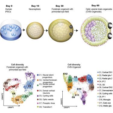
During embryogenesis, optic vesicles develop from the diencephalon via a multistep process of organogenesis. Using induced pluripotent stem cell (iPSC)-derived human brain organoids, we attempted to simplify the complexities and demonstrate formation of forebrain-associated bilateral optic vesicles, cellular diversity, and functionality. Around day 30, brain organoids attempt to assemble optic vesicles, which develop progressively as visible structures within 60 days. These optic vesicle-containing brain organoids (OVB-organoids) constitute a developing optic vesicle’s cellular components, including primitive corneal epithelial and lens-like cells, retinal pigment epithelia, retinal progenitor cells, axon-like projections, and electrically active neuronal networks. OVB-organoids also display synapsin-1, CTIP-positive myelinated cortical neurons, and microglia. Interestingly, various light intensities could trigger photosensitive activity of OVB-organoids, and light sensitivities could be reset after transient photobleaching. Thus, brain organoids have the intrinsic ability to self-organize forebrain-associated primitive sensory structures in a topographically restricted manner and can allow interorgan interaction studies within a single organoid.
中文翻译:
人脑类器官组装功能整合的双侧视囊泡
在胚胎发生过程中,视囊泡通过器官发生的多步骤过程从间脑发育。使用诱导多能干细胞 (iPSC) 衍生的人脑类器官,我们试图简化复杂性并证明前脑相关双侧视囊泡的形成、细胞多样性和功能。第 30 天左右,大脑类器官尝试组装视泡,视泡在 60 天内逐渐发育为可见结构。这些含有视泡的脑类器官 (OVB-organoids) 构成了发育中的视泡的细胞成分,包括原始角膜上皮和晶状体样细胞、视网膜色素上皮细胞、视网膜祖细胞、轴突样突起和电活性神经元网络。OVB 类器官还显示突触蛋白 1、CTIP 阳性有髓皮质神经元、和小胶质细胞。有趣的是,不同的光强度可以触发 OVB 类器官的光敏活性,并且在瞬时光漂白后可以重置光敏度。因此,大脑类器官具有以受地形限制的方式自组织前脑相关原始感觉结构的内在能力,并且可以允许在单个类器官内进行器官间相互作用研究。


 京公网安备 11010802027423号