当前位置:
X-MOL 学术
›
Laser Photonics Rev.
›
论文详情
Our official English website, www.x-mol.net, welcomes your
feedback! (Note: you will need to create a separate account there.)
Intraoperative Label-Free Photoacoustic Histopathology of Clinical Specimens
Laser & Photonics Reviews ( IF 9.8 ) Pub Date : 2021-08-16 , DOI: 10.1002/lpor.202100124 Jin Woo Baik 1 , Hyojin Kim 1 , Myeongjoo Son 2 , Junwon Choi 2 , Kwang Gi Kim 2 , Jeong Heum Baek 3 , Yeon Ho Park 3 , Jungsuk An 3 , Hae Young Choi 4 , Seon Young Ryu 5 , Jin Young Kim 1 , Kyunghee Byun 2 , Chulhong Kim 1
Laser & Photonics Reviews ( IF 9.8 ) Pub Date : 2021-08-16 , DOI: 10.1002/lpor.202100124 Jin Woo Baik 1 , Hyojin Kim 1 , Myeongjoo Son 2 , Junwon Choi 2 , Kwang Gi Kim 2 , Jeong Heum Baek 3 , Yeon Ho Park 3 , Jungsuk An 3 , Hae Young Choi 4 , Seon Young Ryu 5 , Jin Young Kim 1 , Kyunghee Byun 2 , Chulhong Kim 1
Affiliation
|
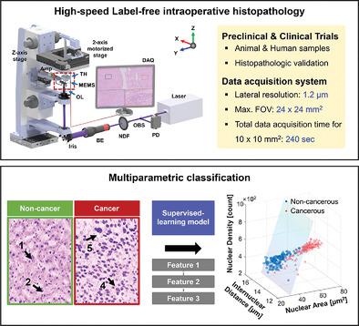
During cancer resection surgeries, intraoperative histopathologic examination of the surgical specimen is crucial for tumor margin identification. A conventional frozen-section analysis requires complex tissue processing, which prolongs surgery and potentially introduces interpretation errors. Here, as a novel approach to label-free intraoperative histopathology, a high-speed reflection-mode ultraviolet photoacoustic microscopy (UV-PAM) system employing a waterproof 1-axis microelectromechanical systems scanner is demonstrated. Label-free nuclear imaging is photoacoustically verified using tissue sections excised from mice and humans. Moreover, by imaging clinical specimens from cancer patients and numerically quantifying the histopathologic results, it is successfully demonstrated that the proposed UV-PAM system has great potential as an alternative intraoperative histopathology method with minimal tissue preparation processes.
中文翻译:

临床标本的术中无标记光声组织病理学
在癌症切除手术中,手术标本的术中组织病理学检查对于识别肿瘤边缘至关重要。传统的冰冻切片分析需要复杂的组织处理,这会延长手术时间并可能引入解释错误。在这里,作为无标记术中组织病理学的一种新方法,展示了一种采用防水 1 轴微机电系统扫描仪的高速反射模式紫外光声显微镜 (UV-PAM) 系统。使用从小鼠和人类切除的组织切片对无标记核成像进行光声验证。此外,通过对癌症患者的临床标本进行成像并对组织病理学结果进行数值量化,
更新日期:2021-10-19
中文翻译:
临床标本的术中无标记光声组织病理学
在癌症切除手术中,手术标本的术中组织病理学检查对于识别肿瘤边缘至关重要。传统的冰冻切片分析需要复杂的组织处理,这会延长手术时间并可能引入解释错误。在这里,作为无标记术中组织病理学的一种新方法,展示了一种采用防水 1 轴微机电系统扫描仪的高速反射模式紫外光声显微镜 (UV-PAM) 系统。使用从小鼠和人类切除的组织切片对无标记核成像进行光声验证。此外,通过对癌症患者的临床标本进行成像并对组织病理学结果进行数值量化,


 京公网安备 11010802027423号