Atherosclerosis ( IF 5.3 ) Pub Date : 2021-08-15 , DOI: 10.1016/j.atherosclerosis.2021.08.027 Ville Rantasalo 1 , Jarmo Gunn 1 , Tuomas Kiviniemi 2 , Jussi Hirvonen 3 , Ilkka Saarenpää 4 , Juri Kivelev 4 , Melissa Rahi 4 , Elli Lassila 5 , Jaakko Rinne 4 , Dan Laukka 4
|
Background and aims
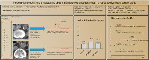
Patients with intracranial aneurysms (IA) have excess mortality for cardiovascular diseases, but little is known on whether atherosclerotic manifestations and IA coexist. We investigated abdominal aortic calcification index (ACI) association with unruptured and ruptured IAs.
Methods
This retrospective case-control study reviews all tertiary centers patients (n = 24,660) who had undergone head computed tomography angiography (CTA), magnetic resonance angiography (MRA) or digital subtraction angiography (DSA) for any reason between January 2003 and May 2018. Patients (n = 2020) with unruptured or ruptured IAs were identified, and patients with available abdominal CT were included. IA patients were matched by sex and age to controls (available abdomen CT, no IAs) in ratio of 1:3. ACI was measured from abdomen CT scans and patient records were reviewed.
Results
1720 patients (216 ruptured IA (rIA), 246 unruptured IA (UIA) and 1258 control) were included. Mean age was 62.9 ± 11.9 years and 58.2% were female. ACI (OR 1.02 per increment, 95%CI 1.01–1.03) and ACI>3 (OR 5.77, 95%CI 3.29–10.11) increased risk for rIA compared to matched controls. UIA patients' ACI was significantly higher but ACI did not increase odds for UIA compared to matched controls. History of coronary artery disease was less frequent in rIA patients. There was no calcification in aorta in 8.8% rIA and 13.6% UIA patients (matched controls 25.7% and 22.6% respectively, p < 0.01).
Conclusions
Aortic calcification is greater in rIA and UIA patients than matched controls. ACI increases risk for rIAs.
中文翻译:
腹主动脉钙化指数预测颅内动脉瘤:回顾性病例对照研究
背景和目标
颅内动脉瘤(IA)患者的心血管疾病死亡率过高,但关于动脉粥样硬化表现和 IA 是否并存尚不清楚。我们调查了腹主动脉钙化指数 (ACI) 与未破裂和破裂 IA 的关系。
方法
这项回顾性病例对照研究回顾了 2003 年 1 月至 2018 年 5 月期间因任何原因接受头颅 CT 血管造影 (CTA)、磁共振血管造影 (MRA) 或数字减影血管造影 (DSA) 的所有三级中心患者 (n = 24,660)。确定了未破裂或破裂 IA 的患者(n = 2020),并纳入了可用腹部 CT 的患者。IA 患者按性别和年龄与对照组(可用腹部 CT,无 IA)以 1:3 的比例匹配。ACI 是通过腹部 CT 扫描测量的,并审查了患者记录。
结果
包括 1720 名患者(216 名 IA 破裂(rIA)、246 名未破裂 IA(UIA)和 1258 名对照)。平均年龄为 62.9 ± 11.9 岁,58.2% 为女性。与匹配的对照相比,ACI(OR 1.02 每个增量,95%CI 1.01–1.03)和 ACI>3(OR 5.77,95%CI 3.29–10.11)增加了 rIA 的风险。UIA 患者的 ACI 显着更高,但与匹配的对照组相比,ACI 并未增加 UIA 的几率。rIA 患者的冠状动脉疾病史较少。8.8% rIA 和 13.6% UIA 患者的主动脉没有钙化(匹配对照分别为 25.7% 和 22.6%,p < 0.01)。
结论
rIA 和 UIA 患者的主动脉钙化程度高于匹配的对照组。ACI 会增加 rIA 的风险。


 京公网安备 11010802027423号