Experimental Neurology ( IF 4.6 ) Pub Date : 2021-08-13 , DOI: 10.1016/j.expneurol.2021.113837 Vito S Hernández 1 , Mario A Zetter 1 , Enrique C Guerra 1 , Ileana Hernández-Araiza 2 , Nikita Karuzin 3 , Oscar R Hernández-Pérez 1 , Lee E Eiden 4 , Limei Zhang 1
|
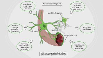
We examined cell type-specific expression and distribution of rat brain angiotensin-converting enzyme 2 (ACE2), the receptor for SARS-CoV-2, in the rodent brain. ACE2 is ubiquitously present in brain vasculature, with the highest density of ACE2 expressing capillaries found in the olfactory bulb, the hypothalamic paraventricular, supraoptic, and mammillary nuclei, the midbrain substantia nigra and ventral tegmental area, and the hindbrain pontine nucleus, the pre-Bötzinger complex, and nucleus of tractus solitarius. ACE2 was expressed in astrocytes and astrocytic foot processes, pericytes and endothelial cells, key components of the blood-brain barrier. We found discrete neuronal groups immunopositive for ACE2 in brainstem respiratory rhythm generating centers, including the pontine nucleus, the parafascicular/retrotrapezoid nucleus, the parabrachial nucleus, the Bötzinger, and pre-Bötzinger complexes and the nucleus of tractus solitarius; in the arousal-related pontine reticular nucleus and gigantocellular reticular nuclei; in brainstem aminergic nuclei, including substantia nigra, ventral tegmental area, dorsal raphe, and locus coeruleus; in the epithalamic habenula, hypothalamic paraventricular and supramammillary nuclei; and in the hippocampus. Identification of ACE2-expressing neurons in rat brain within well-established functional circuits facilitates prediction of possible neurological manifestations of brain ACE2 dysregulation during and after COVID-19 infection.
中文翻译:
大鼠大脑中 ACE2 的表达:对 COVID-19 相关神经系统表现的影响
我们检测了大鼠脑血管紧张素转换酶 2 (ACE2)(SARS-CoV-2 的受体)在啮齿类动物大脑中的细胞类型特异性表达和分布。ACE2普遍存在于脑脉管系统中,在嗅球、下丘脑室旁核、视上核和乳头核、中脑黑质和腹侧被盖区以及后脑桥核、前脑桥核中发现表达ACE2的毛细血管密度最高。 Bötzinger 复合体和孤束核。ACE2 在星形胶质细胞和星形胶质细胞足突、周细胞和内皮细胞(血脑屏障的关键组成部分)中表达。我们发现脑干呼吸节律发生中心的 ACE2 免疫阳性离散神经元组,包括脑桥核、束旁/后梯形核、臂旁核、Bötzinger 和前 Bötzinger 复合体以及孤束核;在与觉醒相关的脑桥网状核和巨细胞网状核中;脑干胺能核,包括黑质、腹侧被盖区、中缝背侧和蓝斑;在上丘脑缰核、下丘脑室旁核和乳头上核中;和在海马体中。


 京公网安备 11010802027423号