当前位置:
X-MOL 学术
›
NMR Biomed.
›
论文详情
Our official English website, www.x-mol.net, welcomes your
feedback! (Note: you will need to create a separate account there.)
Joint total variation-based reconstruction of multiparametric magnetic resonance images for mapping tissue types
NMR in Biomedicine ( IF 2.7 ) Pub Date : 2021-08-13 , DOI: 10.1002/nbm.4597 Shraddha Pandey 1, 2 , A David Snider 2 , Wilfrido A Moreno 2 , Harshan Ravi 1 , Ali Bilgin 3 , Natarajan Raghunand 1, 4
NMR in Biomedicine ( IF 2.7 ) Pub Date : 2021-08-13 , DOI: 10.1002/nbm.4597 Shraddha Pandey 1, 2 , A David Snider 2 , Wilfrido A Moreno 2 , Harshan Ravi 1 , Ali Bilgin 3 , Natarajan Raghunand 1, 4
Affiliation
|
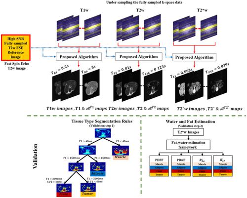
Multispectral analysis of coregistered multiparametric magnetic resonance (MR) images provides a powerful method for tissue phenotyping and segmentation. Acquisition of a sufficiently varied set of multicontrast MR images and parameter maps to objectively define multiple normal and pathologic tissue types can require long scan times. Accelerated MRI on clinical scanners with multichannel receivers exploits techniques such as parallel imaging, while accelerated preclinical MRI scanning must rely on alternate approaches. In this work, tumor-bearing mice were imaged at 7 T to acquire k-space data corresponding to a series of images with varying T1-, T2- and T2*-weighting. A joint reconstruction framework is proposed to reconstruct a series of T1-weighted images and corresponding T1 maps simultaneously from undersampled Cartesian k-space data. The ambiguity introduced by undersampling was resolved by using model-based constraints and structural information from a reference fully sampled image as the joint total variation prior. This process was repeated to reconstruct T2-weighted and T2*-weighted images and corresponding maps of T2 and T2* from undersampled Cartesian k-space data. Validation of the reconstructed images and parameter maps was carried out by computing tissue-type maps, as well as maps of the proton density fat fraction (PDFF), proton density water fraction (PDwF), fat relaxation rate (
 and water relaxation rate (
 from the reconstructed data, and comparing them with ground truth (GT) equivalents. Tissue-type maps computed using 18% k-space data were visually similar to GT tissue-type maps, with dice coefficients ranging from 0.43 to 0.73 for tumor, fluid adipose and muscle tissue types. The mean T1 and T2 values within each tissue type computed using only 18% k-space data were within 8%–10% of the GT values from fully sampled data. The PDFF and PDwF maps computed using 27% k-space data were within 3%–15% of GT values and showed good agreement with the expected values for the four tissue types.
中文翻译:

基于联合总变异的多参数磁共振图像重建用于绘制组织类型
配准多参数磁共振 (MR) 图像的多光谱分析为组织表型和分割提供了一种强大的方法。获取一组足够多的多对比 MR 图像和参数图以客观地定义多种正常和病理组织类型可能需要较长的扫描时间。具有多通道接收器的临床扫描仪上的加速 MRI 利用并行成像等技术,而加速临床前 MRI 扫描必须依赖替代方法。在这项工作中,荷瘤小鼠在 7 T 下成像,以获取与具有不同 T1、T2 和 T2* 加权的一系列图像相对应的 k 空间数据。提出了一种联合重建框架,用于从欠采样的笛卡尔 k 空间数据中同时重建一系列 T1 加权图像和相应的 T1 图。通过使用基于模型的约束和来自参考完全采样图像的结构信息作为联合总变化先验来解决欠采样引入的模糊性。重复此过程以从欠采样的笛卡尔 k 空间数据重建 T2 加权和 T2* 加权图像以及 T2 和 T2* 的相应图。通过计算组织类型图以及质子密度脂肪分数 (PDFF)、质子密度水分数 (PDwF)、脂肪松弛率 ( 重复此过程以从欠采样的笛卡尔 k 空间数据重建 T2 加权和 T2* 加权图像以及 T2 和 T2* 的相应图。通过计算组织类型图以及质子密度脂肪分数 (PDFF)、质子密度水分数 (PDwF)、脂肪松弛率 ( 重复此过程以从欠采样的笛卡尔 k 空间数据重建 T2 加权和 T2* 加权图像以及 T2 和 T2* 的相应图。通过计算组织类型图以及质子密度脂肪分数 (PDFF)、质子密度水分数 (PDwF)、脂肪松弛率 (和水弛豫率(
来自重建数据,并将它们与地面实况 (GT) 等效项进行比较。使用 18% k 空间数据计算的组织类型图在视觉上与 GT 组织类型图相似,骰子系数范围从 0.43 到肿瘤、液体脂肪和肌肉组织类型为 0.73。仅使用 18% k 空间数据计算的每种组织类型内的平均 T1 和 T2 值在完全采样数据的 GT 值的 8%–10% 范围内。PDFF 和 PDwF使用 27% k 空间数据计算的图在 GT 值的 3%–15% 范围内,并且与四种组织类型的预期值显示出良好的一致性。
更新日期:2021-08-13
and water relaxation rate (
from the reconstructed data, and comparing them with ground truth (GT) equivalents. Tissue-type maps computed using 18% k-space data were visually similar to GT tissue-type maps, with dice coefficients ranging from 0.43 to 0.73 for tumor, fluid adipose and muscle tissue types. The mean T1 and T2 values within each tissue type computed using only 18% k-space data were within 8%–10% of the GT values from fully sampled data. The PDFF and PDwF maps computed using 27% k-space data were within 3%–15% of GT values and showed good agreement with the expected values for the four tissue types.
中文翻译:
基于联合总变异的多参数磁共振图像重建用于绘制组织类型
配准多参数磁共振 (MR) 图像的多光谱分析为组织表型和分割提供了一种强大的方法。获取一组足够多的多对比 MR 图像和参数图以客观地定义多种正常和病理组织类型可能需要较长的扫描时间。具有多通道接收器的临床扫描仪上的加速 MRI 利用并行成像等技术,而加速临床前 MRI 扫描必须依赖替代方法。在这项工作中,荷瘤小鼠在 7 T 下成像,以获取与具有不同 T1、T2 和 T2* 加权的一系列图像相对应的 k 空间数据。提出了一种联合重建框架,用于从欠采样的笛卡尔 k 空间数据中同时重建一系列 T1 加权图像和相应的 T1 图。通过使用基于模型的约束和来自参考完全采样图像的结构信息作为联合总变化先验来解决欠采样引入的模糊性。重复此过程以从欠采样的笛卡尔 k 空间数据重建 T2 加权和 T2* 加权图像以及 T2 和 T2* 的相应图。通过计算组织类型图以及质子密度脂肪分数 (PDFF)、质子密度水分数 (PDwF)、脂肪松弛率 ( 重复此过程以从欠采样的笛卡尔 k 空间数据重建 T2 加权和 T2* 加权图像以及 T2 和 T2* 的相应图。通过计算组织类型图以及质子密度脂肪分数 (PDFF)、质子密度水分数 (PDwF)、脂肪松弛率 ( 重复此过程以从欠采样的笛卡尔 k 空间数据重建 T2 加权和 T2* 加权图像以及 T2 和 T2* 的相应图。通过计算组织类型图以及质子密度脂肪分数 (PDFF)、质子密度水分数 (PDwF)、脂肪松弛率 (
和水弛豫率(
来自重建数据,并将它们与地面实况 (GT) 等效项进行比较。使用 18% k 空间数据计算的组织类型图在视觉上与 GT 组织类型图相似,骰子系数范围从 0.43 到肿瘤、液体脂肪和肌肉组织类型为 0.73。仅使用 18% k 空间数据计算的每种组织类型内的平均 T1 和 T2 值在完全采样数据的 GT 值的 8%–10% 范围内。PDFF 和 PDwF使用 27% k 空间数据计算的图在 GT 值的 3%–15% 范围内,并且与四种组织类型的预期值显示出良好的一致性。


 京公网安备 11010802027423号