Archives of Biochemistry and Biophysics ( IF 3.8 ) Pub Date : 2021-08-12 , DOI: 10.1016/j.abb.2021.109003 Miki Kuribayashi 1 , Yusuke Kawaguchi 1 , Hirofumi Teshima 1 , Hisateru Yamaguchi 2 , Hideki Tatsukawa 1 , Kiyotaka Hitomi 1
|
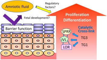
During fetal development, the barrier function of the fetal skin is developed under specific conditions for epidermis formation. In keratinocyte differentiation, the well-orchestrated production and modification of various structural proteins are induced. We assessed the epidermal barrier function in different fetal stages by evaluating the enzymatic activity of cross-linking proteins, transglutaminases, and the permeation of fluorescence dye in the stained epidermal sections. During days 15.5–17.5 in gestation, the enzymatic activities in the epidermis appeared to increase significantly; meanwhile, dye permeation was substantially decreased, suggesting the formation of a protective barrier. For the fetal epidermis formation in the earlier stage, unclarified stimulating factors in the amniotic fluid (AF) are possible to promote barrier function by stimulating keratinocyte differentiation. Thus, we performed proteomic spectrometric (MS) analysis on the components in the AF at different fetal stages. Also, we investigated the promotive ability of the components using a cultured keratinocyte differentiation system. According to the MS analysis, the AF components appeared to exhibit stage-specific variations, where possible unique functions have been identified. We also found that adding the AF from each stage to the medium for cultured keratinocytes specifically enhanced the levels of the differentiation markers. These results provide information on the possible role of AF that contains regulatory factors on keratinocyte differentiation.
中文翻译:
小鼠羊水对不同胎儿阶段角质形成细胞分化刺激能力的研究
在胎儿发育过程中,胎儿皮肤的屏障功能在特定条件下发育,以形成表皮。在角质形成细胞分化中,诱导了各种结构蛋白的精心策划的产生和修饰。我们通过评估染色表皮切片中交联蛋白、转谷氨酰胺酶的酶活性和荧光染料的渗透来评估不同胎儿阶段的表皮屏障功能。在妊娠第 15.5-17.5 天,表皮中的酶活性似乎显着增加;同时,染料渗透性大幅下降,表明形成了保护屏障。对于胎儿表皮形成的早期阶段,羊水(AF)中未明确的刺激因子可能通过刺激角质形成细胞分化来促进屏障功能。因此,我们对不同胎儿阶段 AF 中的成分进行了蛋白质组光谱 (MS) 分析。此外,我们还使用培养的角质形成细胞分化系统研究了成分的促进能力。根据 MS 分析,AF 组件似乎表现出特定阶段的变化,其中可能的独特功能已被识别。我们还发现,将每个阶段的 AF 添加到培养的角质形成细胞的培养基中,可以特异性提高分化标记物的水平。这些结果提供了有关 AF 可能发挥的作用的信息,其中包含对角质形成细胞分化的调节因子。


 京公网安备 11010802027423号