Experimental Eye Research ( IF 3.0 ) Pub Date : 2021-08-11 , DOI: 10.1016/j.exer.2021.108720 Cristina Romo-Valera 1 , Miguel Pérez-Garrastachu 1 , Raquel Hernáez-Moya 1 , Maddalen Rodriguez-Astigarraga 1 , Paula Romano-Ruiz 1 , Jaime Etxebarria 2 , Jon Arluzea 1 , Noelia Andollo 3
|
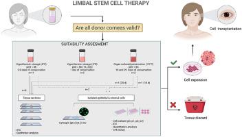
The transplantation of expansions of limbal epithelial stem cells (LESC) remains one of the most efficient therapies for the treatment of limbal stem cell deficiency (LSCD) to date. However, the available donor corneas are scarce, and the corneas conserved for long time, under hypothermic conditions (after 7 days) or in culture (more than 28 days), are usually discarded due to poor viability of the endothelial cells. To establish an objective criterion for the utilisation or discarding of corneas as a source of LESC, we characterized, by immunohistochemistry analysis, donor corneas conserved in different conditions and for different periods of time. We also studied the potency of LESCs isolated from these corneas and maintained in culture up to 3 cell passages. We hoped that the study of markers of LESCs present in both the corneoscleral histological sections and the cell cultures would show the adequacy of the methods used for cell isolation and how fit the LESC enrichment of the obtained cell populations to be expanded was. Thus, the expressions of markers of the cells residing in the human limbal and corneal epithelium (cytokeratin CK15 and CK12, vimentin, Collagen VII, p63α, ABCG2, Ki67, Integrin β4, ZO1, and melan A) were analysed in sections of corneoscleral tissues conserved in hypothermic conditions for 2–9 days with post-mortem time (pmt) < 8 h or for 1 day with pmt > 16 h, and in sclerocorneal rims maintained in an organ culture medium for 29 days. Cell populations isolated from donor corneoscleral tissues were also assessed based on these markers to verify the adequacy of isolation methods and the potential of expanding LESCs from these tissues. Positivity for several putative stem cell markers such as CK15 and p63α was detected in all corneoscleral tissues, although a decrease was recorded in the ones conserved for longer times. The barrier function and the ability to adhere to the extracellular matrix were maintained in all the analysed tissues. In limbal epithelial cell cultures, a simultaneous decrease in the melan A melanocyte marker and the putative stem cell markers was detected, suggesting a close relationship between the melanocytes and the limbal stem cells of the niche. Holoclones stained with putative stem cell markers were obtained from long-term, hypothermic, stored sclerocorneal rims. The results showed that the remaining sclerocorneal rims after corneal transplantation, which were conserved under hypothermic conditions for up to 7 days and would have been discarded at a first glance, still maintained their potential as a source of LESC cultures.
中文翻译:
在不同时间和储存方法后表征角膜作为干样角膜缘上皮细胞的来源
迄今为止,角膜缘上皮干细胞 (LESC) 扩增的移植仍然是治疗角膜缘干细胞缺陷 (LSCD) 的最有效疗法之一。然而,可用的供体角膜稀缺,并且在低温条件下(7天后)或培养(超过28天)下长时间保存的角膜通常由于内皮细胞活力差而被丢弃。为了建立使用或丢弃角膜作为 LESC 来源的客观标准,我们通过免疫组织化学分析表征了在不同条件下和不同时间段保存的供体角膜。我们还研究了从这些角膜中分离出的 LESCs 的效力,并在培养中保持最多 3 次细胞传代。我们希望对角巩膜组织切片和细胞培养物中存在的 LESCs 标记的研究将显示用于细胞分离的方法的充分性以及获得的细胞群的 LESC 富集是如何适合的。因此,在角膜巩膜组织切片中分析了人角膜缘和角膜上皮细胞标记物(细胞角蛋白 CK15 和 CK12、波形蛋白、胶原 VII、p63α、ABCG2、Ki67、整合素 β4、ZO1 和黑色素 A)的表达在低温条件下保存 2-9 天,死后时间 (pmt) < 8 小时或保存 1 天,pmt > 16 小时,并在器官培养基中维持 29 天的巩膜角膜边缘。还基于这些标记评估了从供体角巩膜组织中分离的细胞群,以验证分离方法的充分性以及从这些组织中扩增 LESC 的潜力。在所有角巩膜组织中都检测到了几种推定的干细胞标记物(例如 CK15 和 p63α)的阳性,尽管在保存时间较长的那些组织中记录到了下降。在所有分析的组织中都保持了屏障功能和粘附到细胞外基质的能力。在角膜缘上皮细胞培养物中,检测到黑色素 A 黑色素细胞标志物和推定的干细胞标志物同时减少,表明黑色素细胞和生态位的角膜缘干细胞之间存在密切关系。用推定的干细胞标记物染色的全克隆来自长期、低温、储存的巩膜角膜缘。结果表明,角膜移植后剩余的角膜边缘,在低温条件下保存长达 7 天,乍一看会被丢弃,仍然保持其作为 LESC 培养物来源的潜力。


 京公网安备 11010802027423号