当前位置:
X-MOL 学术
›
Microsc. Res. Tech.
›
论文详情
Our official English website, www.x-mol.net, welcomes your
feedback! (Note: you will need to create a separate account there.)
Automatic cell counting for phase-contrast microscopic images based on a combination of Otsu and watershed segmentation method
Microscopy Research and Technique ( IF 2.0 ) Pub Date : 2021-08-09 , DOI: 10.1002/jemt.23893 Yuefei Lin 1 , Yong Diao 1 , Yongzhao Du 1, 2, 3 , Jianguang Zhang 1 , Ling Li 1 , Peizhong Liu 1, 2, 3
Microscopy Research and Technique ( IF 2.0 ) Pub Date : 2021-08-09 , DOI: 10.1002/jemt.23893 Yuefei Lin 1 , Yong Diao 1 , Yongzhao Du 1, 2, 3 , Jianguang Zhang 1 , Ling Li 1 , Peizhong Liu 1, 2, 3
Affiliation
|
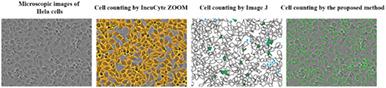
Cell counting plays a vital role in biomedical researches. However, manual cell counting is time-consuming, laborious, and low efficiency and has a high counting error rate problem. An automatic counting approach for Hela cells of phase-contrast microscopic images is proposed based on the combination of Otsu and watershed segmentation methods to solve the mentioned issues. Firstly, image preprocessing is performed. Secondly, the Otsu method was used to obtain an automatic global optimal threshold for segmentation to achieve batch counting of images. Thirdly, the marker watershed was performed to separate adherent cells and to avoid over-segmentation simultaneously. Finally, cells in phase-contrast microscopic images were counted by detecting the numbers of connected domains in the binary image. Taking the manual counting result as the counting standard and MIS, INC, and ACC are used as evaluation indicators. The experimental results showed that the average values of MIS, INC, and ACC of the proposed method are only 3.31%, 3.49%, and 96.69%, respectively. Additionally, each cell image was counted only takes 0.65 s on averagely. To further test the performance of the proposed method, a comparative experiment was carried out by Image J, and the result shows that the proposed method has a better counting performance with a higher average accuracy of 96.55% to Image J with 93.39%.The proposed method for cell counting is simple, feasible, fast and high accurate, and it can be used as an effective method for cell counting of the phase-contrast microscopic images.
中文翻译:

基于Otsu和分水岭分割方法的相衬显微图像自动细胞计数
细胞计数在生物医学研究中起着至关重要的作用。然而,人工细胞计数费时、费力、效率低,且存在计数错误率高的问题。针对上述问题,提出了一种基于Otsu和分水岭分割方法相结合的显微相衬图像Hela细胞自动计数方法。首先,进行图像预处理。其次,利用Otsu方法获得自动全局最优分割阈值,实现图像的批量计数。第三,进行标记分水岭以分离贴壁细胞并同时避免过度分割。最后,通过检测二值图像中连接域的数量,对相差显微图像中的细胞进行计数。以人工计数结果为计数标准,以MIS、INC、ACC为评价指标。实验结果表明,所提方法的MIS、INC和ACC的平均值分别仅为3.31%、3.49%和96.69%。此外,每个细胞图像的计数平均只需要 0.65 秒。为进一步测试所提方法的性能,通过Image J进行对比实验,结果表明所提方法具有更好的计数性能,平均准确率为96.55%,平均准确率为93.39%。细胞计数方法简单、可行、快速、准确度高,可作为相衬显微图像细胞计数的有效方法。实验结果表明,所提方法的MIS、INC和ACC的平均值分别仅为3.31%、3.49%和96.69%。此外,每个细胞图像的计数平均只需要 0.65 秒。为进一步测试所提方法的性能,通过Image J进行对比实验,结果表明所提方法具有更好的计数性能,平均准确率为96.55%,平均准确率为93.39%。细胞计数方法简单、可行、快速、准确度高,可作为相衬显微图像细胞计数的有效方法。实验结果表明,所提方法的MIS、INC和ACC的平均值分别仅为3.31%、3.49%和96.69%。此外,每个细胞图像的计数平均只需要 0.65 秒。为进一步测试所提方法的性能,通过Image J进行对比实验,结果表明所提方法具有更好的计数性能,平均准确率为96.55%,平均准确率为93.39%。细胞计数方法简单、可行、快速、准确度高,可作为相衬显微图像细胞计数的有效方法。平均 65 秒。为进一步测试所提方法的性能,通过Image J进行对比实验,结果表明所提方法具有更好的计数性能,平均准确率为96.55%,平均准确率为93.39%。细胞计数方法简单、可行、快速、准确度高,可作为相衬显微图像细胞计数的有效方法。平均 65 秒。为进一步测试所提方法的性能,通过Image J进行对比实验,结果表明所提方法具有更好的计数性能,平均准确率为96.55%,平均准确率为93.39%。细胞计数方法简单、可行、快速、准确度高,可作为相衬显微图像细胞计数的有效方法。
更新日期:2021-08-09
中文翻译:
基于Otsu和分水岭分割方法的相衬显微图像自动细胞计数
细胞计数在生物医学研究中起着至关重要的作用。然而,人工细胞计数费时、费力、效率低,且存在计数错误率高的问题。针对上述问题,提出了一种基于Otsu和分水岭分割方法相结合的显微相衬图像Hela细胞自动计数方法。首先,进行图像预处理。其次,利用Otsu方法获得自动全局最优分割阈值,实现图像的批量计数。第三,进行标记分水岭以分离贴壁细胞并同时避免过度分割。最后,通过检测二值图像中连接域的数量,对相差显微图像中的细胞进行计数。以人工计数结果为计数标准,以MIS、INC、ACC为评价指标。实验结果表明,所提方法的MIS、INC和ACC的平均值分别仅为3.31%、3.49%和96.69%。此外,每个细胞图像的计数平均只需要 0.65 秒。为进一步测试所提方法的性能,通过Image J进行对比实验,结果表明所提方法具有更好的计数性能,平均准确率为96.55%,平均准确率为93.39%。细胞计数方法简单、可行、快速、准确度高,可作为相衬显微图像细胞计数的有效方法。实验结果表明,所提方法的MIS、INC和ACC的平均值分别仅为3.31%、3.49%和96.69%。此外,每个细胞图像的计数平均只需要 0.65 秒。为进一步测试所提方法的性能,通过Image J进行对比实验,结果表明所提方法具有更好的计数性能,平均准确率为96.55%,平均准确率为93.39%。细胞计数方法简单、可行、快速、准确度高,可作为相衬显微图像细胞计数的有效方法。实验结果表明,所提方法的MIS、INC和ACC的平均值分别仅为3.31%、3.49%和96.69%。此外,每个细胞图像的计数平均只需要 0.65 秒。为进一步测试所提方法的性能,通过Image J进行对比实验,结果表明所提方法具有更好的计数性能,平均准确率为96.55%,平均准确率为93.39%。细胞计数方法简单、可行、快速、准确度高,可作为相衬显微图像细胞计数的有效方法。平均 65 秒。为进一步测试所提方法的性能,通过Image J进行对比实验,结果表明所提方法具有更好的计数性能,平均准确率为96.55%,平均准确率为93.39%。细胞计数方法简单、可行、快速、准确度高,可作为相衬显微图像细胞计数的有效方法。平均 65 秒。为进一步测试所提方法的性能,通过Image J进行对比实验,结果表明所提方法具有更好的计数性能,平均准确率为96.55%,平均准确率为93.39%。细胞计数方法简单、可行、快速、准确度高,可作为相衬显微图像细胞计数的有效方法。


 京公网安备 11010802027423号