当前位置:
X-MOL 学术
›
Eur. J. Neurosci.
›
论文详情
Our official English website, www.x-mol.net, welcomes your
feedback! (Note: you will need to create a separate account there.)
Calibrating rhythmic stimulation parameters to individual electroencephalography markers: The consistency of individual alpha frequency in practical lab settings
European Journal of Neuroscience ( IF 2.7 ) Pub Date : 2021-08-07 , DOI: 10.1111/ejn.15418 Shanice E W Janssens 1, 2 , Alexander T Sack 1, 2, 3, 4 , Sanne Ten Oever 1, 5, 6 , Tom A de Graaf 1, 2, 4
European Journal of Neuroscience ( IF 2.7 ) Pub Date : 2021-08-07 , DOI: 10.1111/ejn.15418 Shanice E W Janssens 1, 2 , Alexander T Sack 1, 2, 3, 4 , Sanne Ten Oever 1, 5, 6 , Tom A de Graaf 1, 2, 4
Affiliation
|
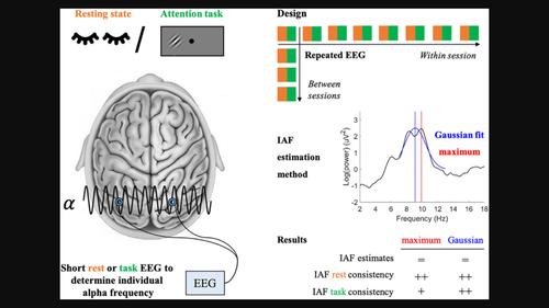
Rhythmic stimulation can be applied to modulate neuronal oscillations. Such ‘entrainment’ is optimized when stimulation frequency is individually calibrated based on magneto/encephalography markers. It remains unknown how consistent such individual markers are across days/sessions, within a session, or across cognitive states, hemispheres and estimation methods, especially in a realistic, practical, lab setting. We here estimated individual alpha frequency (IAF) repeatedly from short electroencephalography (EEG) measurements at rest or during an attention task (cognitive state), using single parieto-occipital electrodes in 24 participants on 4 days (between-sessions), with multiple measurements over an hour on 1 day (within-session). First, we introduce an algorithm to automatically reject power spectra without a sufficiently clear peak to ensure unbiased IAF estimations. Then we estimated IAF via the traditional ‘maximum’ method and a ‘Gaussian fit’ method. IAF was reliable within- and between-sessions for both cognitive states and hemispheres, though task-IAF estimates tended to be more variable. Overall, the ‘Gaussian fit’ method was more reliable than the ‘maximum’ method. Furthermore, we evaluated how far from an approximated ‘true’ task-related IAF the selected ‘stimulation frequency’ was, when calibrating this frequency based on a short rest-EEG, a short task-EEG, or simply selecting 10 Hz for all participants. For the ‘maximum’ method, rest-EEG calibration was best, followed by task-EEG, and then 10 Hz. For the ‘Gaussian fit’ method, rest-EEG and task-EEG-based calibration were similarly accurate, and better than 10 Hz. These results lead to concrete recommendations about valid, and automated, estimation of individual oscillation markers in experimental and clinical settings.
中文翻译:

根据个体脑电图标记校准节律性刺激参数:实际实验室环境中个体 α 频率的一致性
节奏刺激可用于调节神经元振荡。当基于磁/脑电图标记单独校准刺激频率时,这种“夹带”得到优化。目前尚不清楚这些个体标记在不同天/会话、会话内或跨认知状态、半球和估计方法之间的一致性如何,尤其是在现实、实用的实验室环境中。我们在这里通过短时间脑电图 (EEG) 测量在休息或注意力任务期间(认知状态)重复估计个体 α 频率 (IAF),在 4 天(会话之间)使用 24 名参与者的单个顶枕电极,进行多次测量1 天(会期内)超过一个小时。第一的,我们引入了一种算法来自动拒绝没有足够清晰的峰值的功率谱,以确保无偏的 IAF 估计。然后我们通过传统的“最大”方法和“高斯拟合”方法估计 IAF。IAF 在认知状态和大脑半球的会话内和会话之间都是可靠的,尽管任务 IAF 估计往往更具可变性。总体而言,“高斯拟合”方法比“最大”方法更可靠。此外,我们评估了在基于短休息脑电图、短任务脑电图或简单地为所有参与者选择 10 赫兹来校准这个频率时,所选的“刺激频率”与近似的“真实”任务相关 IAF 有多远. 对于“最大”方法,休息脑电图校准最好,其次是任务脑电图,然后是 10 赫兹。对于“高斯拟合”方法,静息脑电图和基于任务脑电图的校准同样准确,并且优于 10 Hz。这些结果导致了关于在实验和临床环境中对个体振荡标记进行有效和自动化估计的具体建议。
更新日期:2021-08-07
中文翻译:
根据个体脑电图标记校准节律性刺激参数:实际实验室环境中个体 α 频率的一致性
节奏刺激可用于调节神经元振荡。当基于磁/脑电图标记单独校准刺激频率时,这种“夹带”得到优化。目前尚不清楚这些个体标记在不同天/会话、会话内或跨认知状态、半球和估计方法之间的一致性如何,尤其是在现实、实用的实验室环境中。我们在这里通过短时间脑电图 (EEG) 测量在休息或注意力任务期间(认知状态)重复估计个体 α 频率 (IAF),在 4 天(会话之间)使用 24 名参与者的单个顶枕电极,进行多次测量1 天(会期内)超过一个小时。第一的,我们引入了一种算法来自动拒绝没有足够清晰的峰值的功率谱,以确保无偏的 IAF 估计。然后我们通过传统的“最大”方法和“高斯拟合”方法估计 IAF。IAF 在认知状态和大脑半球的会话内和会话之间都是可靠的,尽管任务 IAF 估计往往更具可变性。总体而言,“高斯拟合”方法比“最大”方法更可靠。此外,我们评估了在基于短休息脑电图、短任务脑电图或简单地为所有参与者选择 10 赫兹来校准这个频率时,所选的“刺激频率”与近似的“真实”任务相关 IAF 有多远. 对于“最大”方法,休息脑电图校准最好,其次是任务脑电图,然后是 10 赫兹。对于“高斯拟合”方法,静息脑电图和基于任务脑电图的校准同样准确,并且优于 10 Hz。这些结果导致了关于在实验和临床环境中对个体振荡标记进行有效和自动化估计的具体建议。


 京公网安备 11010802027423号