当前位置:
X-MOL 学术
›
Microsc. Res. Tech.
›
论文详情
Our official English website, www.x-mol.net, welcomes your
feedback! (Note: you will need to create a separate account there.)
Developmental morphological analyses on the preglottal salivary gland in Japanese quails (Coturnix japonica)
Microscopy Research and Technique ( IF 2.0 ) Pub Date : 2021-08-03 , DOI: 10.1002/jemt.23892 Mahmoud Osman Khalifa 1, 2 , Mahmoud Abd-Elkareem 3 , Wafaa Gaber 3 , Tao-Sheng Li 2 , Abdelmohaimen Mostafa Saleh 3
Microscopy Research and Technique ( IF 2.0 ) Pub Date : 2021-08-03 , DOI: 10.1002/jemt.23892 Mahmoud Osman Khalifa 1, 2 , Mahmoud Abd-Elkareem 3 , Wafaa Gaber 3 , Tao-Sheng Li 2 , Abdelmohaimen Mostafa Saleh 3
Affiliation
|
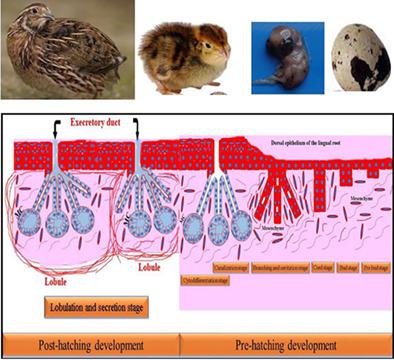
To understand the development of the mucous preglottal salivary gland in Coturnix japonica (Japanese quail), morphological and histochemical studies were performed on 20 healthy Japanese quail embryos (aging from 10th to 17th incubation days) and 25 healthy quail chicks (aging from 0th to 60th days). The primordia of preglottal salivary gland were observed as an epithelial bud at the early embryonic stage, which then elongated and differentiated into secretory units by the end of this stage. In Japanese quails, the preglottal salivary gland was a mucous polystomatic tubuloalveolar unpaired gland composed of two lateral portions and a middle one embedded into the submucosa of the lingual root. The gland openings accompanied taste pore (8.17 μm) of taste buds associated salivary glands type; some skeletal muscle fibers embedded among secretory lobules extended from muscle cricohyoideus at 14th day-old quail chick. Also, both herbts corpuscles and secretory motor plexus could be detected among secretory lobules. Based on our investigations, the development of the preglottal salivary gland could clearly be distinguished in the embryonic stage into pre bud and bud stages at 10th day old, cord and branching stages ended by cavitation at 11th day old, canalization stage at 13th day old, lobulation and secretory stages by the 17th day old. The secretory materials showed different histochemical reactions ended with highly alcinophilic mucous indicated highly sialomucin (acidic) content. Myoepithelial cells could be demonstrated at a 17-day old quail embryo and thereafter surrounded the secretory endpieces of the preglottal salivary gland.
中文翻译:

日本鹌鹑(Coturnix japonica)声门前唾液腺发育形态学分析
为了了解Coturnix japonica(日本鹌鹑)的声门前粘液唾液腺的发育,对 20 名健康日本人进行了形态学和组织化学研究。鹌鹑胚胎(从第 10 天到第 17 天孵化)和 25 只健康鹌鹑小鸡(从第 0 天到第 60 天老化)。声门前唾液腺原基在胚胎早期被观察为上皮芽,然后在该阶段结束时伸长并分化为分泌单位。在日本鹌鹑中,声门前唾液腺是一个粘液多口小管肺泡未配对腺体,由两个侧面部分和一个嵌入舌根粘膜下层的中间部分组成。腺体开口伴随着味蕾相关唾液腺型的味孔(8.17 μm);14 日龄的鹌鹑雏鸡从环舌骨肌延伸出的分泌小叶中嵌入了一些骨骼肌纤维。此外,在分泌小叶中可以检测到草药小体和分泌运动神经丛。根据我们的调查,声门前唾液腺的发育在胚胎阶段可以清楚地区分为前芽和 10 日龄的芽期,11 日龄以空化结束的索和分枝期,13 日龄的管化阶段,到第 17 天大的分叶和分泌阶段。分泌物显示出不同的组织化学反应,以高嗜酸性粘液结束,表明高唾液酸粘蛋白(酸性)含量。肌上皮细胞可以在一个 17 天大的鹌鹑胚胎中得到证实,然后围绕在声门前唾液腺的分泌末端。13 日龄时为管化期,17 日龄时为分叶和分泌期。分泌物显示出不同的组织化学反应,以高嗜酸性粘液结束,表明高唾液酸粘蛋白(酸性)含量。肌上皮细胞可以在一个 17 天大的鹌鹑胚胎中得到证实,然后围绕在声门前唾液腺的分泌末端。13 日龄时为管化期,17 日龄时为分叶和分泌期。分泌物显示出不同的组织化学反应,以高嗜酸性粘液结束,表明高唾液酸粘蛋白(酸性)含量。肌上皮细胞可以在一个 17 天大的鹌鹑胚胎中得到证实,然后围绕在声门前唾液腺的分泌末端。
更新日期:2021-08-03
中文翻译:
日本鹌鹑(Coturnix japonica)声门前唾液腺发育形态学分析
为了了解Coturnix japonica(日本鹌鹑)的声门前粘液唾液腺的发育,对 20 名健康日本人进行了形态学和组织化学研究。鹌鹑胚胎(从第 10 天到第 17 天孵化)和 25 只健康鹌鹑小鸡(从第 0 天到第 60 天老化)。声门前唾液腺原基在胚胎早期被观察为上皮芽,然后在该阶段结束时伸长并分化为分泌单位。在日本鹌鹑中,声门前唾液腺是一个粘液多口小管肺泡未配对腺体,由两个侧面部分和一个嵌入舌根粘膜下层的中间部分组成。腺体开口伴随着味蕾相关唾液腺型的味孔(8.17 μm);14 日龄的鹌鹑雏鸡从环舌骨肌延伸出的分泌小叶中嵌入了一些骨骼肌纤维。此外,在分泌小叶中可以检测到草药小体和分泌运动神经丛。根据我们的调查,声门前唾液腺的发育在胚胎阶段可以清楚地区分为前芽和 10 日龄的芽期,11 日龄以空化结束的索和分枝期,13 日龄的管化阶段,到第 17 天大的分叶和分泌阶段。分泌物显示出不同的组织化学反应,以高嗜酸性粘液结束,表明高唾液酸粘蛋白(酸性)含量。肌上皮细胞可以在一个 17 天大的鹌鹑胚胎中得到证实,然后围绕在声门前唾液腺的分泌末端。13 日龄时为管化期,17 日龄时为分叶和分泌期。分泌物显示出不同的组织化学反应,以高嗜酸性粘液结束,表明高唾液酸粘蛋白(酸性)含量。肌上皮细胞可以在一个 17 天大的鹌鹑胚胎中得到证实,然后围绕在声门前唾液腺的分泌末端。13 日龄时为管化期,17 日龄时为分叶和分泌期。分泌物显示出不同的组织化学反应,以高嗜酸性粘液结束,表明高唾液酸粘蛋白(酸性)含量。肌上皮细胞可以在一个 17 天大的鹌鹑胚胎中得到证实,然后围绕在声门前唾液腺的分泌末端。


 京公网安备 11010802027423号