当前位置:
X-MOL 学术
›
Angew. Chem. Int. Ed.
›
论文详情
Our official English website, www.x-mol.net, welcomes your
feedback! (Note: you will need to create a separate account there.)
Hard X-Ray Nanotomography for 3D Analysis of Coking in Nickel-Based Catalysts
Angewandte Chemie International Edition ( IF 16.1 ) Pub Date : 2021-08-02 , DOI: 10.1002/anie.202106380 Sebastian Weber 1, 2 , Darren Batey 3 , Silvia Cipiccia 4 , Matthias Stehle 1 , Ken L Abel 5 , Roger Gläser 5 , Thomas L Sheppard 1, 2
Angewandte Chemie International Edition ( IF 16.1 ) Pub Date : 2021-08-02 , DOI: 10.1002/anie.202106380 Sebastian Weber 1, 2 , Darren Batey 3 , Silvia Cipiccia 4 , Matthias Stehle 1 , Ken L Abel 5 , Roger Gläser 5 , Thomas L Sheppard 1, 2
Affiliation
|
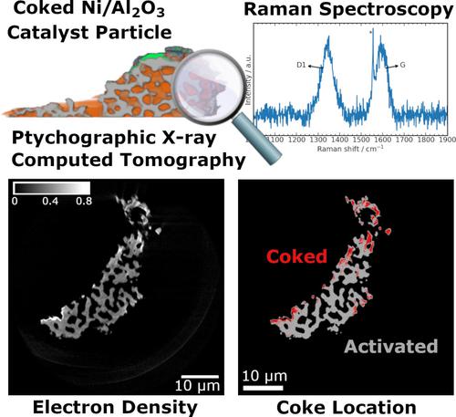
Understanding catalyst deactivation by coking is crucial for knowledge-based catalyst and process design in reactions with carbonaceous species. Post-mortem analysis of catalyst coking is often performed by bulk characterization methods. Here, hard X-ray ptychographic computed tomography (PXCT) was used to study Ni/Al2O3 catalysts for CO2 methanation and CH4 dry reforming after artificial coking treatment. PXCT generated quantitative 3D maps of local electron density at ca. 80 nm resolution, allowing to visualize and evaluate the severity of coking in entire catalyst particles of ca. 40 μm diameter. Coking was primarily revealed in the nanoporous solid, which was not detectable in resolved macropores. Coke formation was independently confirmed by operando Raman spectroscopy. PXCT is highlighted as an emerging characterization tool for nanoscale identification, co-localization, and potentially quantification of deactivation phenomena in 3D space within entire catalyst particles.
中文翻译:

用于镍基催化剂结焦 3D 分析的硬 X 射线纳米断层扫描
了解焦化导致的催化剂失活对于碳质反应中基于知识的催化剂和工艺设计至关重要。催化剂结焦的事后分析通常通过本体表征方法进行。本文采用硬X射线叠层计算机断层扫描(PXCT)研究了人工焦化处理后用于CO 2甲烷化和CH 4干重整的Ni/Al 2 O 3催化剂。 PXCT 生成了约 1 处局部电子密度的定量 3D 图。 80 nm 分辨率,可以可视化并评估整个催化剂颗粒中焦化的严重程度。直径40微米。结焦主要出现在纳米多孔固体中,而在解析的大孔中无法检测到结焦。焦炭的形成通过操作拉曼光谱独立证实。 PXCT 被强调为一种新兴的表征工具,用于纳米级识别、共定位以及整个催化剂颗粒内 3D 空间失活现象的潜在量化。
更新日期:2021-09-20
中文翻译:
用于镍基催化剂结焦 3D 分析的硬 X 射线纳米断层扫描
了解焦化导致的催化剂失活对于碳质反应中基于知识的催化剂和工艺设计至关重要。催化剂结焦的事后分析通常通过本体表征方法进行。本文采用硬X射线叠层计算机断层扫描(PXCT)研究了人工焦化处理后用于CO 2甲烷化和CH 4干重整的Ni/Al 2 O 3催化剂。 PXCT 生成了约 1 处局部电子密度的定量 3D 图。 80 nm 分辨率,可以可视化并评估整个催化剂颗粒中焦化的严重程度。直径40微米。结焦主要出现在纳米多孔固体中,而在解析的大孔中无法检测到结焦。焦炭的形成通过操作拉曼光谱独立证实。 PXCT 被强调为一种新兴的表征工具,用于纳米级识别、共定位以及整个催化剂颗粒内 3D 空间失活现象的潜在量化。


 京公网安备 11010802027423号