当前位置:
X-MOL 学术
›
FEBS Open Bio
›
论文详情
Our official English website, www.x-mol.net, welcomes your
feedback! (Note: you will need to create a separate account there.)
RIP3-mediated necroptosis increases neuropathic pain via microglia activation: necrostatin-1 has therapeutic potential
FEBS Open Bio ( IF 2.8 ) Pub Date : 2021-07-28 , DOI: 10.1002/2211-5463.13258 Ping Fang 1 , Gangqiang Sun 1 , Jingyu Wang 1
FEBS Open Bio ( IF 2.8 ) Pub Date : 2021-07-28 , DOI: 10.1002/2211-5463.13258 Ping Fang 1 , Gangqiang Sun 1 , Jingyu Wang 1
Affiliation
|
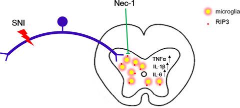
Neuropathic pain (NP) is a clinical symptom that accompanies many diseases. We investigated the effect of receptor-interacting protein kinase 3 (RIP3)-regulated necroptosis on NP and explored its relationship with microglia, in order to provide a theoretical basis for further research and provide new insights into the treatment of NP. In this study, the spared nerve injury (SNI) model was used along with intervention with necrostatin and the inhibitor of necroptosis necrostatin-1 (Nec-1). Pain behavior tests were performed 1 and 3 days before the nerve injury (or sham) operation, and on days 1, 3, 5, 7, 10, and 14 after the operation. The spinal cord tissues were collected for detection of RIP3 expression and distribution, changes in the number of microglia cells, activation of necroptosis, and the level of proinflammatory factors. Collected spinal cord tissues were analyzed using western blot, immunohistochemistry, immunofluorescence, immunoprecipitation assays, and ELISA, respectively. We found that, compared with the sham group, the expression of RIP3 protein in the spinal cord of rats in the SNI group increased from 3 to 14 days after surgery. Immunofluorescence staining showed that RIP3 was coexpressed with the microglia and the number of microglia increased significantly in the SNI model group. The results of immunoprecipitation assays suggested that a RIP3-mediated necroptosis pathway promotes NP. After treatment with Nec-1, the expression of RIP3 protein and the number of microglia were significantly reduced, and the expression levels of TNF-α, IL-1β, and IL-6 in spinal dorsal horns were significantly decreased. These results indicate that RIP3 promotes necroptosis to increase the occurrence of NP via microglia.
中文翻译:

RIP3 介导的坏死性凋亡通过小胶质细胞激活增加神经性疼痛:necrostatin-1 具有治疗潜力
神经性疼痛(NP)是伴随许多疾病的临床症状。我们研究了受体相互作用蛋白激酶3(RIP3)调节的坏死性凋亡对NP的影响,并探讨了其与小胶质细胞的关系,以期为进一步研究提供理论基础,并为NP的治疗提供新的见解。在本研究中,保留神经损伤 (SNI) 模型与 necrostatin 和 necrostatin-1 (necrostatin-1) 抑制剂 (Nec-1) 一起使用。疼痛行为测试在神经损伤(或假手术)前 1 天和 3 天,以及术后第 1、3、5、7、10 和 14 天进行。采集脊髓组织检测RIP3的表达和分布、小胶质细胞数量的变化、坏死性凋亡的激活情况以及促炎因子的水平。分别使用蛋白质印迹、免疫组织化学、免疫荧光、免疫沉淀测定和 ELISA 分析收集的脊髓组织。我们发现,与假手术组相比,SNI组大鼠脊髓中RIP3蛋白的表达在术后3~14天增加。免疫荧光染色显示RIP3与小胶质细胞共表达,SNI模型组小胶质细胞数量明显增加。免疫沉淀测定的结果表明,RIP3 介导的坏死性凋亡途径促进了 NP。Nec-1处理后RIP3蛋白表达和小胶质细胞数量明显减少,脊髓背角TNF-α、IL-1β、IL-6表达水平明显降低。
更新日期:2021-10-02
中文翻译:
RIP3 介导的坏死性凋亡通过小胶质细胞激活增加神经性疼痛:necrostatin-1 具有治疗潜力
神经性疼痛(NP)是伴随许多疾病的临床症状。我们研究了受体相互作用蛋白激酶3(RIP3)调节的坏死性凋亡对NP的影响,并探讨了其与小胶质细胞的关系,以期为进一步研究提供理论基础,并为NP的治疗提供新的见解。在本研究中,保留神经损伤 (SNI) 模型与 necrostatin 和 necrostatin-1 (necrostatin-1) 抑制剂 (Nec-1) 一起使用。疼痛行为测试在神经损伤(或假手术)前 1 天和 3 天,以及术后第 1、3、5、7、10 和 14 天进行。采集脊髓组织检测RIP3的表达和分布、小胶质细胞数量的变化、坏死性凋亡的激活情况以及促炎因子的水平。分别使用蛋白质印迹、免疫组织化学、免疫荧光、免疫沉淀测定和 ELISA 分析收集的脊髓组织。我们发现,与假手术组相比,SNI组大鼠脊髓中RIP3蛋白的表达在术后3~14天增加。免疫荧光染色显示RIP3与小胶质细胞共表达,SNI模型组小胶质细胞数量明显增加。免疫沉淀测定的结果表明,RIP3 介导的坏死性凋亡途径促进了 NP。Nec-1处理后RIP3蛋白表达和小胶质细胞数量明显减少,脊髓背角TNF-α、IL-1β、IL-6表达水平明显降低。


 京公网安备 11010802027423号