当前位置:
X-MOL 学术
›
NMR Biomed.
›
论文详情
Our official English website, www.x-mol.net, welcomes your
feedback! (Note: you will need to create a separate account there.)
Tiny golden angle ultrashort echo-time lung imaging in mice
NMR in Biomedicine ( IF 2.7 ) Pub Date : 2021-07-28 , DOI: 10.1002/nbm.4591 Anke Balasch 1 , Patrick Metze 1 , Hao Li 2, 3 , Wolfgang Rottbauer 1 , Alireza Abaei 3 , Volker Rasche 1, 3
NMR in Biomedicine ( IF 2.7 ) Pub Date : 2021-07-28 , DOI: 10.1002/nbm.4591 Anke Balasch 1 , Patrick Metze 1 , Hao Li 2, 3 , Wolfgang Rottbauer 1 , Alireza Abaei 3 , Volker Rasche 1, 3
Affiliation
|
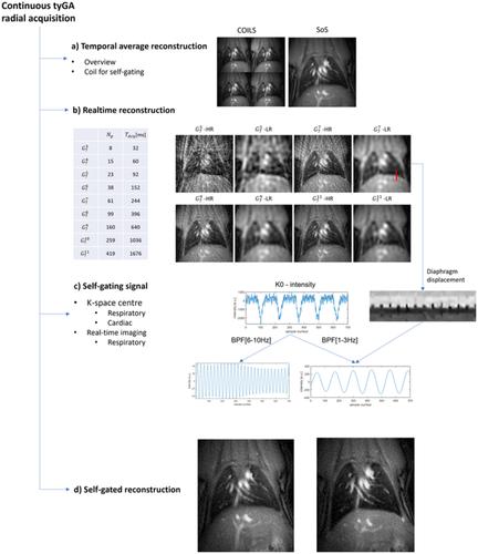
Imaging the lung parenchyma with MRI is particularly difficult in small animals due to the high respiratory and heart rates, and ultrashort T2* at high magnetic field strength caused by the high susceptibilities induced by the air–tissue interfaces. In this study, a 2D ultrashort echo-time (UTE) technique was combined with tiny golden angle (tyGA) ordering. Data were acquired continuously at 11.7 T and retrospective center-of-k-space gating was applied to reconstruct respiratory multistage images. Lung (proton) density (fP), T2*, signal-to-noise ratio (SNR), fractional ventilation (FV) and perfusion (f) were quantified, and the application to dynamic contrast agent (CA)-enhanced (DCE) qualitative perfusion assessment tested. The interobserver and intraobserver and interstudy reproducibility of the quantitative parameters were investigated. High-quality images of the lung parenchyma could be acquired in all animals. Over all lung regions a mean T2* of 0.20 ± 0.05 ms was observed. FV resulted as 0.31 ± 0.13, and a trend towards lower SNR values during inspiration (EX: SNR = 12.48 ± 6.68, IN: SNR = 11.79 ± 5.86) and a significant (P < 0.001) decrease in lung density (EX: fP = 0.69 ± 0.13, IN: fP = 0.62 ± 0.13) were observed. Quantitative perfusion results as 34.63 ± 9.05 mL/cm3/min (systole) and 32.77 ± 8.55 mL/cm3/min (diastole) on average. The CA dynamics could be assessed and, because of the continuous nature of the data acquisition, reconstructed at different temporal resolutions. Where a good to excellent interobserver reproducibility and an excellent intraobserver reproducibility resulted, the interstudy reproducibility was only fair to good. In conclusion, the combination of tiny golden angles with UTE (2D tyGA UTE) resulted in a reliable imaging technique for lung morphology and function in mice, providing uniform k-space coverage and thus low-artefact images of the lung parenchyma after gating.
中文翻译:

小鼠微小金角超短回波时间肺部成像
由于呼吸和心率高,以及由空气 - 组织界面引起的高磁化率引起的高磁场强度下的超短 T2*,在小动物中使用 MRI 对肺实质进行成像特别困难。在这项研究中,2D 超短回波时间 (UTE) 技术与微小黄金角 (tyGA) 排序相结合。在 11.7 T 下连续采集数据,并应用回顾性 k 空间中心门控来重建呼吸多阶段图像。肺(质子)密度 ( f P )、T2*、信噪比 (SNR)、分数通气 (FV) 和灌注 ( f) 进行了量化,并测试了动态造影剂 (CA) 增强 (DCE) 定性灌注评估的应用。研究了定量参数的观察者间和观察者内以及研究间的可重复性。所有动物都可以获得高质量的肺实质图像。在所有肺区域中,观察到平均 T2* 为 0.20 ± 0.05 ms。FV 为 0.31 ± 0.13,吸气时 SNR 值趋于降低(EX:SNR = 12.48 ± 6.68,IN:SNR = 11.79 ± 5.86) ,肺密度显着降低( P < 0.001)(EX: f P = 0.69 ± 0.13, IN: f P = 0.62 ± 0.13) 被观察到。定量灌注结果为 34.63 ± 9.05 mL/cm 3/min(收缩期)和平均 32.77 ± 8.55 mL/cm 3 /min(舒张期)。可以评估 CA 动态,并且由于数据采集的连续性,可以在不同的时间分辨率下重建。如果产生了良好到极好的观察者间重现性和良好的观察者内重现性,则研究间重现性只是一般到好。总之,微小的黄金角与 UTE (2D tyGA UTE) 的结合为小鼠的肺形态和功能提供了一种可靠的成像技术,提供了均匀的 k 空间覆盖,从而提供了门控后肺实质的低伪影图像。
更新日期:2021-10-06
中文翻译:
小鼠微小金角超短回波时间肺部成像
由于呼吸和心率高,以及由空气 - 组织界面引起的高磁化率引起的高磁场强度下的超短 T2*,在小动物中使用 MRI 对肺实质进行成像特别困难。在这项研究中,2D 超短回波时间 (UTE) 技术与微小黄金角 (tyGA) 排序相结合。在 11.7 T 下连续采集数据,并应用回顾性 k 空间中心门控来重建呼吸多阶段图像。肺(质子)密度 ( f P )、T2*、信噪比 (SNR)、分数通气 (FV) 和灌注 ( f) 进行了量化,并测试了动态造影剂 (CA) 增强 (DCE) 定性灌注评估的应用。研究了定量参数的观察者间和观察者内以及研究间的可重复性。所有动物都可以获得高质量的肺实质图像。在所有肺区域中,观察到平均 T2* 为 0.20 ± 0.05 ms。FV 为 0.31 ± 0.13,吸气时 SNR 值趋于降低(EX:SNR = 12.48 ± 6.68,IN:SNR = 11.79 ± 5.86) ,肺密度显着降低( P < 0.001)(EX: f P = 0.69 ± 0.13, IN: f P = 0.62 ± 0.13) 被观察到。定量灌注结果为 34.63 ± 9.05 mL/cm 3/min(收缩期)和平均 32.77 ± 8.55 mL/cm 3 /min(舒张期)。可以评估 CA 动态,并且由于数据采集的连续性,可以在不同的时间分辨率下重建。如果产生了良好到极好的观察者间重现性和良好的观察者内重现性,则研究间重现性只是一般到好。总之,微小的黄金角与 UTE (2D tyGA UTE) 的结合为小鼠的肺形态和功能提供了一种可靠的成像技术,提供了均匀的 k 空间覆盖,从而提供了门控后肺实质的低伪影图像。


 京公网安备 11010802027423号