当前位置:
X-MOL 学术
›
Microsc. Res. Tech.
›
论文详情
Our official English website, www.x-mol.net, welcomes your
feedback! (Note: you will need to create a separate account there.)
Ultrastructural characterization of the intestine of the Eurasian common moorhen using scanning electron microscopy and light microscopy
Microscopy Research and Technique ( IF 2.0 ) Pub Date : 2021-07-28 , DOI: 10.1002/jemt.23888 Basma G Hanafy 1 , Mohamed M A Abumandour 1 , Ramadan Kandyle 2 , Naglaa F Bassuoni 1
Microscopy Research and Technique ( IF 2.0 ) Pub Date : 2021-07-28 , DOI: 10.1002/jemt.23888 Basma G Hanafy 1 , Mohamed M A Abumandour 1 , Ramadan Kandyle 2 , Naglaa F Bassuoni 1
Affiliation
|
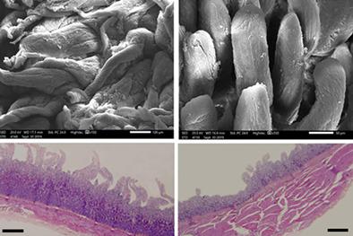
The present investigation focused on the morphological features of the intestine of Eurasian common moorhen by the aid of scanning electron microscopy and histological, morphometric, and statistical examinations. The intestinal villi were varied in shape along the intestinal tract; the duodenal villi were elongated and twisted, the jejunal villi were tongue-like, the ileal villi were cuboidal, and the cecal villi were tongue-like and finger-like at the base and body of the cecum. While at the apex of the cecum, it appeared as corrugated longitudinal folds and the rectal villi appeared as columns. The microvilli were present as projections on the surface of villi of the small intestine and the base of the cecum. While at the body and the apex of the cecum, the microvilli appeared as threads and as brush hairs on the rectal villi. The duodenal, jejunal, ileal, cecal, and rectal villi were lined by simple columnar epithelium with goblet cells. The submucosal layer of small intestine consisted of connective tissue fibers along the intestinal tract, but it was absent at the base of the cecum. The tunica musculosa consisted of single longitudinal layer of smooth muscle fibers in the duodenum and rectum. While in the jejunum, ileum, and cecum, it was composed of single circular layer of smooth muscle fibers. The thickness of the tunics differed among the intestinal parts.
中文翻译:

使用扫描电子显微镜和光学显微镜对欧亚普通海鸡肠道的超微结构表征
目前的调查重点是通过扫描电子显微镜和组织学、形态测量学和统计检查的帮助,研究欧亚普通海雀肠道的形态特征。肠绒毛沿肠道形状各异;十二指肠绒毛伸长扭曲,空肠绒毛呈舌状,回肠绒毛呈立方形,盲肠绒毛在盲肠基部和体部呈舌状和指状。而在盲肠顶端,则表现为波纹状纵向皱襞,直肠绒毛表现为柱状。微绒毛作为小肠绒毛表面和盲肠基部的突起存在。而在身体和盲肠顶端,微绒毛在直肠绒毛上表现为线和刷毛。十二指肠,空肠、回肠、盲肠和直肠绒毛的内衬是带有杯状细胞的简单柱状上皮。小肠黏膜下层由沿肠道的结缔组织纤维组成,但在盲肠基部不存在。肌肉膜由十二指肠和直肠中的单层纵向平滑肌纤维组成。而在空肠、回肠和盲肠中,它由单层圆形平滑肌纤维组成。外衣的厚度因肠道部位而异。肌肉膜由十二指肠和直肠中的单层纵向平滑肌纤维组成。而在空肠、回肠和盲肠中,它由单层圆形平滑肌纤维组成。外衣的厚度因肠道部位而异。肌肉膜由十二指肠和直肠中的单层纵向平滑肌纤维组成。而在空肠、回肠和盲肠中,它由单层圆形平滑肌纤维组成。外衣的厚度因肠道部位而异。
更新日期:2021-07-28
中文翻译:
使用扫描电子显微镜和光学显微镜对欧亚普通海鸡肠道的超微结构表征
目前的调查重点是通过扫描电子显微镜和组织学、形态测量学和统计检查的帮助,研究欧亚普通海雀肠道的形态特征。肠绒毛沿肠道形状各异;十二指肠绒毛伸长扭曲,空肠绒毛呈舌状,回肠绒毛呈立方形,盲肠绒毛在盲肠基部和体部呈舌状和指状。而在盲肠顶端,则表现为波纹状纵向皱襞,直肠绒毛表现为柱状。微绒毛作为小肠绒毛表面和盲肠基部的突起存在。而在身体和盲肠顶端,微绒毛在直肠绒毛上表现为线和刷毛。十二指肠,空肠、回肠、盲肠和直肠绒毛的内衬是带有杯状细胞的简单柱状上皮。小肠黏膜下层由沿肠道的结缔组织纤维组成,但在盲肠基部不存在。肌肉膜由十二指肠和直肠中的单层纵向平滑肌纤维组成。而在空肠、回肠和盲肠中,它由单层圆形平滑肌纤维组成。外衣的厚度因肠道部位而异。肌肉膜由十二指肠和直肠中的单层纵向平滑肌纤维组成。而在空肠、回肠和盲肠中,它由单层圆形平滑肌纤维组成。外衣的厚度因肠道部位而异。肌肉膜由十二指肠和直肠中的单层纵向平滑肌纤维组成。而在空肠、回肠和盲肠中,它由单层圆形平滑肌纤维组成。外衣的厚度因肠道部位而异。


 京公网安备 11010802027423号