当前位置:
X-MOL 学术
›
Hum. Brain Mapp.
›
论文详情
Our official English website, www.x-mol.net, welcomes your
feedback! (Note: you will need to create a separate account there.)
Topologically convergent and divergent morphological gray matter networks in early-stage Parkinson's disease with and without mild cognitive impairment
Human Brain Mapping ( IF 3.5 ) Pub Date : 2021-07-28 , DOI: 10.1002/hbm.25606 Xueling Suo 1, 2, 3 , Du Lei 1, 2, 3, 4 , Nannan Li 5 , Junying Li 5 , Jiaxin Peng 5 , Wenbin Li 1, 2, 3 , Jing Yang 1, 2, 3 , Kun Qin 1, 2, 3 , Graham J Kemp 6 , Rong Peng 5 , Qiyong Gong 1, 2, 3
Human Brain Mapping ( IF 3.5 ) Pub Date : 2021-07-28 , DOI: 10.1002/hbm.25606 Xueling Suo 1, 2, 3 , Du Lei 1, 2, 3, 4 , Nannan Li 5 , Junying Li 5 , Jiaxin Peng 5 , Wenbin Li 1, 2, 3 , Jing Yang 1, 2, 3 , Kun Qin 1, 2, 3 , Graham J Kemp 6 , Rong Peng 5 , Qiyong Gong 1, 2, 3
Affiliation
|
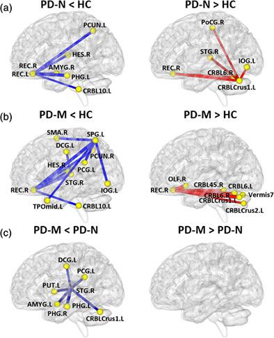
Patients with Parkinson's disease with mild cognitive impairment (PD-M) progress to dementia more frequently than those with normal cognition (PD-N), but the underlying neurobiology remains unclear. This study aimed to define the specific morphological brain network alterations in PD-M, and explore their potential diagnostic value. Twenty-four PD-M patients, 17 PD-N patients, and 29 healthy controls (HC) underwent a structural MRI scan. Similarity between interregional gray matter volume distributions was used to construct individual morphological brain networks. These were analyzed using graph theory and network-based statistics (NBS), and their relationship to neuropsychological tests was assessed. Support vector machine (SVM) was used to perform individual classification. Globally, compared with HC, PD-M showed increased local efficiency (p = .001) in their morphological networks, while PD-N showed decreased normalized path length (p = .008). Locally, similar nodal deficits were found in the rectus and lingual gyrus, and cerebellum of both PD groups relative to HC; additionally in PD-M nodal deficits involved several frontal and parietal regions, correlated with cognitive scores. NBS found that similar connections were involved in the default mode and cerebellar networks of both PD groups (to a greater extent in PD-M), while PD-M, but not PD-N, showed altered connections involving the frontoparietal network. Using connections identified by NBS, SVM allowed discrimination with high accuracy between PD-N and HC (90%), PD-M and HC (85%), and between the two PD groups (65%). These results suggest that default mode and cerebellar disruption characterizes PD, more so in PD-M, whereas frontoparietal disruption has diagnostic potential.
中文翻译:

伴有或不伴有轻度认知障碍的早期帕金森病的拓扑收敛和发散形态灰质网络
患有轻度认知障碍(PD-M)的帕金森病患者比认知正常(PD-N)的帕金森病患者更容易进展为痴呆,但其潜在的神经生物学仍不清楚。本研究旨在明确PD-M中特定的形态学脑网络改变,并探讨其潜在的诊断价值。 24 名 PD-M 患者、17 名 PD-N 患者和 29 名健康对照 (HC) 接受了结构 MRI 扫描。区域间灰质体积分布之间的相似性被用来构建个体形态大脑网络。使用图论和基于网络的统计(NBS)对这些进行分析,并评估它们与神经心理学测试的关系。支持向量机(SVM)用于执行个体分类。在全球范围内,与 HC 相比,PD-M 在其形态网络中显示出局部效率提高 ( p = .001),而 PD-N 显示归一化路径长度缩短 ( p = .008)。就局部而言,与 HC 相比,两个 PD 组的直肌、舌回和小脑均发现类似的淋巴结缺损;此外,PD-M 中的淋巴结缺陷涉及多个额叶和顶叶区域,与认知评分相关。 NBS 发现,两个 PD 组的默认模式和小脑网络中都涉及类似的连接(PD-M 中更严重),而 PD-M(而非 PD-N)显示涉及额顶叶网络的连接发生了改变。使用 NBS 识别的连接,SVM 可以高精度地区分 PD-N 和 HC (90%)、PD-M 和 HC (85%) 以及两个 PD 组 (65%)。这些结果表明,默认模式和小脑破坏是 PD 的特征,在 PD-M 中更是如此,而额顶叶破坏具有诊断潜力。
更新日期:2021-09-19
中文翻译:
伴有或不伴有轻度认知障碍的早期帕金森病的拓扑收敛和发散形态灰质网络
患有轻度认知障碍(PD-M)的帕金森病患者比认知正常(PD-N)的帕金森病患者更容易进展为痴呆,但其潜在的神经生物学仍不清楚。本研究旨在明确PD-M中特定的形态学脑网络改变,并探讨其潜在的诊断价值。 24 名 PD-M 患者、17 名 PD-N 患者和 29 名健康对照 (HC) 接受了结构 MRI 扫描。区域间灰质体积分布之间的相似性被用来构建个体形态大脑网络。使用图论和基于网络的统计(NBS)对这些进行分析,并评估它们与神经心理学测试的关系。支持向量机(SVM)用于执行个体分类。在全球范围内,与 HC 相比,PD-M 在其形态网络中显示出局部效率提高 ( p = .001),而 PD-N 显示归一化路径长度缩短 ( p = .008)。就局部而言,与 HC 相比,两个 PD 组的直肌、舌回和小脑均发现类似的淋巴结缺损;此外,PD-M 中的淋巴结缺陷涉及多个额叶和顶叶区域,与认知评分相关。 NBS 发现,两个 PD 组的默认模式和小脑网络中都涉及类似的连接(PD-M 中更严重),而 PD-M(而非 PD-N)显示涉及额顶叶网络的连接发生了改变。使用 NBS 识别的连接,SVM 可以高精度地区分 PD-N 和 HC (90%)、PD-M 和 HC (85%) 以及两个 PD 组 (65%)。这些结果表明,默认模式和小脑破坏是 PD 的特征,在 PD-M 中更是如此,而额顶叶破坏具有诊断潜力。


 京公网安备 11010802027423号