当前位置:
X-MOL 学术
›
Mol. Pharmaceutics
›
论文详情
Our official English website, www.x-mol.net, welcomes your
feedback! (Note: you will need to create a separate account there.)
Positron Emission Tomography Imaging of Autotaxin in Thyroid and Breast Cancer Models Using [18F]PRIMATX
Molecular Pharmaceutics ( IF 4.5 ) Pub Date : 2021-07-28 , DOI: 10.1021/acs.molpharmaceut.1c00265 Marcus Litchfield 1, 2 , Melinda Wuest 1 , Daryl Glubrecht 1 , Emmanuelle Briard 3 , Yves P Auberson 3 , Todd P W McMullen 1, 2 , David N Brindley 2, 4 , Frank Wuest 1, 2
Molecular Pharmaceutics ( IF 4.5 ) Pub Date : 2021-07-28 , DOI: 10.1021/acs.molpharmaceut.1c00265 Marcus Litchfield 1, 2 , Melinda Wuest 1 , Daryl Glubrecht 1 , Emmanuelle Briard 3 , Yves P Auberson 3 , Todd P W McMullen 1, 2 , David N Brindley 2, 4 , Frank Wuest 1, 2
Affiliation
|
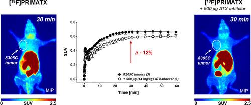
Autotaxin (ATX) is a secreted enzyme responsible for producing lysophosphatidic acid (LPA). The ATX/LPA signaling axis is typically activated in wound healing and tissue repair processes. The ATX/LPA axis is highjacked and upregulated in the progression and persistence of several chronic inflammatory diseases, including cancer. As ATX inhibitors are now progressing to clinical testing, innovative diagnostic tools such as positron emission tomography (PET) are needed to measure ATX expression in vivo accurately. The radiotracer, [18F]PRIMATX, was recently developed and tested for PET imaging of ATX in vivo in a murine melanoma model. The goal of the present work was to further validate [18F]PRIMATX as a PET imaging agent by analyzing its in vivo metabolic stability and suitability for PET imaging of ATX in models of human 8305C thyroid tumor and murine 4T1 breast cancer. [18F]PRIMATX displayed favorable metabolic stability in vivo (65% of intact radiotracer after 60 min p.i.) and provided sufficient tumor uptake profiles in both tumor models. Radiotracer uptake could be blocked by 8–12% in 8305C thyroid tumors in the presence of ATX inhibitor AE-32-NZ70 as determined by PET and ex vivo biodistribution analyses. [18F]PRIMATX also showed high brain uptake, which was reduced by 50% through the administration of ATX inhibitor AE-32-NZ70. [18F]PRIMATX is a suitable radiotracer for PET imaging of ATX in the brain and peripheral tumor tissues.
中文翻译:

使用 [18F]PRIMATX 对甲状腺和乳腺癌模型中的 Autotaxin 进行正电子发射断层扫描成像
Autotaxin (ATX) 是一种分泌酶,负责产生溶血磷脂酸 (LPA)。ATX/LPA 信号轴通常在伤口愈合和组织修复过程中被激活。ATX/LPA 轴在包括癌症在内的几种慢性炎症性疾病的进展和持续存在中被劫持和上调。随着 ATX 抑制剂现在进入临床测试,需要创新的诊断工具,如正电子发射断层扫描 (PET) 来准确测量体内 ATX 的表达。放射性示踪剂 [ 18 F]PRIMATX 是最近开发的,并在小鼠黑色素瘤模型中测试了体内 ATX 的 PET 成像。目前工作的目标是进一步验证 [ 18F]PRIMATX 作为 PET 显像剂,通过分析其在人 8305C 甲状腺肿瘤和鼠 4T1 乳腺癌模型中的体内代谢稳定性和 ATX PET 显像的适用性。[ 18 F]PRIMATX 在体内表现出良好的代谢稳定性(注射 60 分钟后完整的放射性示踪剂的 65%),并在两种肿瘤模型中提供了足够的肿瘤摄取曲线。根据 PET 和离体生物分布分析确定,在存在 ATX 抑制剂 AE-32-NZ70 的情况下,8305C 甲状腺肿瘤中的放射性示踪剂摄取可被阻断 8-12%。[ 18 F]PRIMATX 还表现出高脑摄取,通过 ATX 抑制剂 AE-32-NZ70 的给药降低了 50%。[ 18 F]PRIMATX 是一种适用于脑和外周肿瘤组织中 ATX 的 PET 成像的放射性示踪剂。
更新日期:2021-09-06
中文翻译:
使用 [18F]PRIMATX 对甲状腺和乳腺癌模型中的 Autotaxin 进行正电子发射断层扫描成像
Autotaxin (ATX) 是一种分泌酶,负责产生溶血磷脂酸 (LPA)。ATX/LPA 信号轴通常在伤口愈合和组织修复过程中被激活。ATX/LPA 轴在包括癌症在内的几种慢性炎症性疾病的进展和持续存在中被劫持和上调。随着 ATX 抑制剂现在进入临床测试,需要创新的诊断工具,如正电子发射断层扫描 (PET) 来准确测量体内 ATX 的表达。放射性示踪剂 [ 18 F]PRIMATX 是最近开发的,并在小鼠黑色素瘤模型中测试了体内 ATX 的 PET 成像。目前工作的目标是进一步验证 [ 18F]PRIMATX 作为 PET 显像剂,通过分析其在人 8305C 甲状腺肿瘤和鼠 4T1 乳腺癌模型中的体内代谢稳定性和 ATX PET 显像的适用性。[ 18 F]PRIMATX 在体内表现出良好的代谢稳定性(注射 60 分钟后完整的放射性示踪剂的 65%),并在两种肿瘤模型中提供了足够的肿瘤摄取曲线。根据 PET 和离体生物分布分析确定,在存在 ATX 抑制剂 AE-32-NZ70 的情况下,8305C 甲状腺肿瘤中的放射性示踪剂摄取可被阻断 8-12%。[ 18 F]PRIMATX 还表现出高脑摄取,通过 ATX 抑制剂 AE-32-NZ70 的给药降低了 50%。[ 18 F]PRIMATX 是一种适用于脑和外周肿瘤组织中 ATX 的 PET 成像的放射性示踪剂。


 京公网安备 11010802027423号