Journal of Comparative Pathology ( IF 0.8 ) Pub Date : 2021-07-28 , DOI: 10.1016/j.jcpa.2021.06.004 Kumiko Kimura 1 , Tohru Yanase 2 , Tomoko Kato 2
|
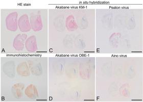
Akabane, Aino and Peaton viruses are closely related arthropod-borne viruses in the genus Orthobunyavirus of the family Peribunyaviridae that can cause congenital abnormalities in cattle, sheep and goats. East Asian Akabane virus strains are subdivided into genogroups Ⅰ and Ⅱ, and the former can also cause non-suppurative encephalomyelitis in post-natal animals. Specific detection of the infecting virus in tissues is essential for accurate diagnosis. Immunohistochemistry (IHC) has been used to identify viral antigen but cannot always detect specific viruses due to potential cross-reactivity of the primary antisera. We compared in-situ hybridization (ISH), based on the use of cocktail probe sets targeted at the RNA of each virus, with IHC for the detection of the specific viruses in tissues of suckling rats inoculated intracerebrally with Akabane (KM-1 or OBE-1 strains), Aino or Peaton viruses at 3 or 7 days of age. Most inoculated rats developed severe neurological signs and histopathological brain lesions including necrosis, spongy degeneration and non-suppurative inflammation. A rabbit polyclonal antiserum immunolabelled antigen of all three viruses within the lesions, whereas ISH specifically detected RNA of each individual virus. The distribution of viral RNA was comparable to that of viral antigens, but tended to be more widespread, especially in immature nervous tissue. Viral antigen and RNA were detected in skeletal muscle and heart of the rats infected with the KM-1 strain of Akabane virus but not with any of the other viruses. This study demonstrates the value of ISH detection of these viruses in a rat model and may prove useful for clarification of the pathogenesis of post-natal arbovirus infection.
中文翻译:
实验感染赤羽基因组Ⅰ和Ⅱ、Aino 和Peaton 病毒的乳鼠的组织病理学、免疫组织化学和原位杂交结果
Akabane、Aino 和 Peaton 病毒是Peribunyaviridae科Orthobunyavirus属中密切相关的节肢动物传播病毒这会导致牛、绵羊和山羊的先天性异常。东亚赤羽病毒株细分为基因组Ⅰ和Ⅱ,前者也可引起出生后动物的非化脓性脑脊髓炎。在组织中特异性检测感染病毒对于准确诊断至关重要。免疫组织化学 (IHC) 已被用于识别病毒抗原,但由于初级抗血清的潜在交叉反应性,无法始终检测到特定病毒。我们比较了原位杂交 (ISH),基于使用针对每种病毒的 RNA 的鸡尾酒探针组,与 IHC 用于检测脑内接种 Akabane(KM-1 或 OBE)的乳鼠组织中的特定病毒-1 株)、Aino 或 Peaton 病毒在 3 或 7 日龄时。大多数接种的大鼠出现严重的神经系统症状和组织病理学脑损伤,包括坏死、海绵状变性和非化脓性炎症。病变内所有三种病毒的兔多克隆抗血清免疫标记抗原,而 ISH 特异性检测每种病毒的 RNA。病毒RNA的分布与病毒抗原的分布相当,但往往更广泛,尤其是在未成熟的神经组织中。在感染 KM-1 赤羽病毒株的大鼠的骨骼肌和心脏中检测到病毒抗原和 RNA,但没有检测到任何其他病毒。该研究证明了在大鼠模型中对这些病毒进行 ISH 检测的价值,并可能有助于阐明出生后虫媒病毒感染的发病机制。海绵状变性和非化脓性炎症。病变内所有三种病毒的兔多克隆抗血清免疫标记抗原,而 ISH 特异性检测每种病毒的 RNA。病毒RNA的分布与病毒抗原的分布相当,但往往更广泛,尤其是在未成熟的神经组织中。在感染 KM-1 赤羽病毒株的大鼠的骨骼肌和心脏中检测到病毒抗原和 RNA,但没有检测到任何其他病毒。该研究证明了在大鼠模型中对这些病毒进行 ISH 检测的价值,并可能有助于阐明出生后虫媒病毒感染的发病机制。海绵状变性和非化脓性炎症。病变内所有三种病毒的兔多克隆抗血清免疫标记抗原,而 ISH 特异性检测每种病毒的 RNA。病毒RNA的分布与病毒抗原的分布相当,但往往更广泛,尤其是在未成熟的神经组织中。在感染 KM-1 赤羽病毒株的大鼠的骨骼肌和心脏中检测到病毒抗原和 RNA,但没有检测到任何其他病毒。该研究证明了在大鼠模型中对这些病毒进行 ISH 检测的价值,并可能有助于阐明出生后虫媒病毒感染的发病机制。而ISH专门检测每个病毒的RNA。病毒RNA的分布与病毒抗原的分布相当,但往往更广泛,尤其是在未成熟的神经组织中。在感染 KM-1 赤羽病毒株的大鼠的骨骼肌和心脏中检测到病毒抗原和 RNA,但没有检测到任何其他病毒。该研究证明了在大鼠模型中对这些病毒进行 ISH 检测的价值,并可能有助于阐明出生后虫媒病毒感染的发病机制。而ISH专门检测每个病毒的RNA。病毒RNA的分布与病毒抗原的分布相当,但往往更广泛,尤其是在未成熟的神经组织中。在感染 KM-1 赤羽病毒株的大鼠的骨骼肌和心脏中检测到病毒抗原和 RNA,但没有检测到任何其他病毒。该研究证明了在大鼠模型中对这些病毒进行 ISH 检测的价值,并可能有助于阐明出生后虫媒病毒感染的发病机制。在感染 KM-1 赤羽病毒株的大鼠的骨骼肌和心脏中检测到病毒抗原和 RNA,但没有检测到任何其他病毒。该研究证明了在大鼠模型中对这些病毒进行 ISH 检测的价值,并可能有助于阐明出生后虫媒病毒感染的发病机制。在感染 KM-1 赤羽病毒株的大鼠的骨骼肌和心脏中检测到病毒抗原和 RNA,但没有检测到任何其他病毒。该研究证明了在大鼠模型中对这些病毒进行 ISH 检测的价值,并可能有助于阐明出生后虫媒病毒感染的发病机制。


 京公网安备 11010802027423号