当前位置:
X-MOL 学术
›
Brain Pathol.
›
论文详情
Our official English website, www.x-mol.net, welcomes your feedback! (Note: you will need to create a separate account there.)
An integrative histopathological and epigenetic characterization of primary intracranial mesenchymal tumors, FET:CREB-fused broadening the spectrum of tumor entities in comparison with their soft tissue counterparts
Brain Pathology ( IF 6.4 ) Pub Date : 2021-07-27 , DOI: 10.1111/bpa.13010 Arnault Tauziède-Espariat 1, 2 , Philipp Sievers 3, 4 , Frédérique Larousserie 5 , Joseph Benzakoun 2, 6 , Delphine Guillemot 7, 8 , Gaëlle Pierron 7, 8 , Mathilde Duchesne 9 , Emmanuelle Uro-Coste 10, 11, 12 , Alexandre Roux 2, 13 , Alexandre Vasiljevic 14 , Tanguy Fenouil 14 , David Meyronet 14 , Karima Mokhtari 15 , Marc Polivka 16 , Audrey Rousseau 17 , Frédérique Bost-Bezeaud 18 , Samir Akoury 19 , Johan Pallud 2, 13 , Chiara Benevello 13 , Lauren Hasty 1 , Albane Gareton 1 , Emmanuèle Lechapt 1 , Fabrice Chrétien 1 , Thomas Blauwblomme 20 , Kévin Beccaria 20 , Stéphanie Puget 20 , Felix Sahm 3, 4, 21 , Pascale Varlet 1, 2 ,
Brain Pathology ( IF 6.4 ) Pub Date : 2021-07-27 , DOI: 10.1111/bpa.13010 Arnault Tauziède-Espariat 1, 2 , Philipp Sievers 3, 4 , Frédérique Larousserie 5 , Joseph Benzakoun 2, 6 , Delphine Guillemot 7, 8 , Gaëlle Pierron 7, 8 , Mathilde Duchesne 9 , Emmanuelle Uro-Coste 10, 11, 12 , Alexandre Roux 2, 13 , Alexandre Vasiljevic 14 , Tanguy Fenouil 14 , David Meyronet 14 , Karima Mokhtari 15 , Marc Polivka 16 , Audrey Rousseau 17 , Frédérique Bost-Bezeaud 18 , Samir Akoury 19 , Johan Pallud 2, 13 , Chiara Benevello 13 , Lauren Hasty 1 , Albane Gareton 1 , Emmanuèle Lechapt 1 , Fabrice Chrétien 1 , Thomas Blauwblomme 20 , Kévin Beccaria 20 , Stéphanie Puget 20 , Felix Sahm 3, 4, 21 , Pascale Varlet 1, 2 ,
Affiliation
|
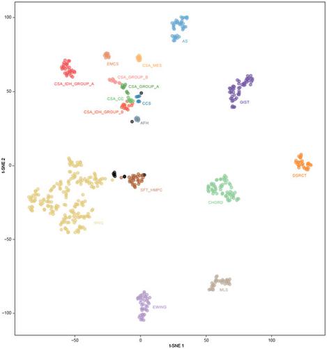
FET:CREB fusions have been described in a variety of tumors from various phenotypes. Recently, these fusion transcripts were reported in intracranial tumors, variably named intracranial mesenchymal myxoid tumors or angiomatoid fibrous histiocytomas. Controversy remains concerning the terminology for these tumors. Here, we report 11 cases of central nervous system mesenchymal tumors with proven FET:CREB fusion. Most DNA methylation profiles were not classifiable using the Heidelberg Brain Tumor or Sarcoma Classifier (v11b4/v12.2). However, by using unsupervised t-SNE and hierarchical clustering analyses, six of the cases constituted a distinct cluster. The remaining four tumors showed no obvious relation to any of the other referenced classes but were close to the clusters of extra-CNS angiomatoid fibrous histiocytomas (n = 1), clear cell sarcomas (n = 1), or solitary fibrous tumors (n = 2). Our findings confirm that intracranial FET:CREB-fused tumors do not represent a single molecular tumor entity, although most samples clustered close to each other, indicating the existence of a distinct epigenetic group that could potentially be partially masked by the low number of cases included. Further analyses are needed to characterize intracranial FET:CREB fused-defined tumors in more detail.
中文翻译:

原发性颅内间充质肿瘤的综合组织病理学和表观遗传学特征,与软组织对应物相比,FET:CREB 融合拓宽了肿瘤实体的范围
FET:CREB 融合已在来自不同表型的多种肿瘤中得到描述。最近,在颅内肿瘤中报道了这些融合转录物,这些肿瘤被命名为颅内间充质粘液样肿瘤或血管瘤样纤维组织细胞瘤。关于这些肿瘤的术语仍然存在争议。在这里,我们报告了 11 例证实 FET:CREB 融合的中枢神经系统间充质肿瘤。大多数 DNA 甲基化谱无法使用海德堡脑肿瘤或肉瘤分类器 (v11b4/v12.2) 进行分类。然而,通过使用无监督的 t-SNE 和层次聚类分析,其中六个案例构成了一个不同的集群。其余四个肿瘤与任何其他参考类别均无明显关系,但接近中枢神经系统外血管瘤样纤维组织细胞瘤簇(n = 1),透明细胞肉瘤(n = 1)或孤立性纤维瘤(n = 2)。我们的研究结果证实,颅内 FET:CREB 融合肿瘤并不代表单个分子肿瘤实体,尽管大多数样本彼此聚集在一起,表明存在一个独特的表观遗传组,该组可能被少数病例部分掩盖. 需要进一步分析以更详细地表征颅内 FET:CREB 融合定义的肿瘤。
更新日期:2021-07-27
中文翻译:
原发性颅内间充质肿瘤的综合组织病理学和表观遗传学特征,与软组织对应物相比,FET:CREB 融合拓宽了肿瘤实体的范围
FET:CREB 融合已在来自不同表型的多种肿瘤中得到描述。最近,在颅内肿瘤中报道了这些融合转录物,这些肿瘤被命名为颅内间充质粘液样肿瘤或血管瘤样纤维组织细胞瘤。关于这些肿瘤的术语仍然存在争议。在这里,我们报告了 11 例证实 FET:CREB 融合的中枢神经系统间充质肿瘤。大多数 DNA 甲基化谱无法使用海德堡脑肿瘤或肉瘤分类器 (v11b4/v12.2) 进行分类。然而,通过使用无监督的 t-SNE 和层次聚类分析,其中六个案例构成了一个不同的集群。其余四个肿瘤与任何其他参考类别均无明显关系,但接近中枢神经系统外血管瘤样纤维组织细胞瘤簇(n = 1),透明细胞肉瘤(n = 1)或孤立性纤维瘤(n = 2)。我们的研究结果证实,颅内 FET:CREB 融合肿瘤并不代表单个分子肿瘤实体,尽管大多数样本彼此聚集在一起,表明存在一个独特的表观遗传组,该组可能被少数病例部分掩盖. 需要进一步分析以更详细地表征颅内 FET:CREB 融合定义的肿瘤。


 京公网安备 11010802027423号