当前位置:
X-MOL 学术
›
NMR Biomed.
›
论文详情
Our official English website, www.x-mol.net, welcomes your
feedback! (Note: you will need to create a separate account there.)
Differentiation of symptomatic and asymptomatic carotid intraplaque hemorrhage using 3D high-resolution diffusion-weighted stack of stars imaging
NMR in Biomedicine ( IF 2.7 ) Pub Date : 2021-07-23 , DOI: 10.1002/nbm.4582 Seong-Eun Kim 1, 2 , Dennis L Parker 1, 2 , John A Roberts 1, 2 , Gerald S Treiman 3 , Matthew Alexander 1, 2 , Hediyeh Baradaran 1, 2 , Adam de Havenon 4 , J Scott McNally 1, 2
NMR in Biomedicine ( IF 2.7 ) Pub Date : 2021-07-23 , DOI: 10.1002/nbm.4582 Seong-Eun Kim 1, 2 , Dennis L Parker 1, 2 , John A Roberts 1, 2 , Gerald S Treiman 3 , Matthew Alexander 1, 2 , Hediyeh Baradaran 1, 2 , Adam de Havenon 4 , J Scott McNally 1, 2
Affiliation
|
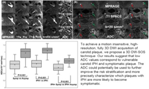
Ischemic events related to carotid disease are far more strongly associated with plaque instability than stenosis. 3D high-resolution diffusion-weighted (DW) imaging can provide quantitative diffusion measurements on carotid atherosclerosis and may improve detection of vulnerable intraplaque hemorrhage (IPH). The 3D DW-stack of stars (SOS) sequence was implemented with 3D SOS acquisition combined with DW preparation. After simulation of signals created from 3D DW-SOS, phantom studies were performed. Three healthy subjects and 20 patients with carotid disease were recruited. Apparent diffusion coefficient (ADC) values were statistically analyzed on three subgroups by using a two-group comparison Wilcoxon–Mann–Whitney U test with p values less than 0.05: symptomatic versus asymptomatic; IPH-positive versus IPH-negative; and IPH-positive symptomatic versus asymptomatic plaques to determine the relationship with plaque vulnerability. ADC values calculated by 3D DW-SOS provided values similar to those calculated from other techniques. Mean ADC of symptomatic plaque was significantly lower than asymptomatic plaque (0.68 ± 0.18 vs. 0.98 ± 0.16 x 10⁻³ mm2/s, p < 0.001). ADC was also significantly lower in IPH-positive versus IPH-negative plaque (0.68 ± 0.13 vs. 1.04 ± 0.11 x 10⁻³ mm2/s, p < 0.001). Additionally, ADC was significantly lower in symptomatic versus asymptomatic IPH-positive plaque (0.57 ± 0.09 vs. 0.75 ± 0.11 x 10⁻³ mm2/s, p < 0.001). Our results provide strong evidence that ADC measurements from 3D DW-SOS correlate with the symptomatic status of extracranial internal carotid artery plaque. Further, ADC improved discrimination of symptomatic plaque in IPH. These data suggest that diffusion characteristics may improve detection of destabilized plaque leading to elevated stroke risk.
中文翻译:

使用 3D 高分辨率扩散加权星成像技术区分有症状和无症状的颈动脉斑块内出血
与颈动脉疾病相关的缺血事件与斑块不稳定的相关性比与狭窄的相关性要强得多。 3D 高分辨率扩散加权 (DW) 成像可以提供颈动脉粥样硬化的定量扩散测量,并可以改善易损斑块内出血 (IPH) 的检测。 3D DW 星堆 (SOS) 序列是通过 3D SOS 采集与 DW 准备相结合来实现的。对 3D DW-SOS 创建的信号进行模拟后,进行了模型研究。招募了 3 名健康受试者和 20 名颈动脉疾病患者。通过使用两组比较 Wilcoxon-Mann-Whitney U 检验对三个亚组的表观扩散系数 (ADC) 值进行统计分析, p值小于 0.05:有症状与无症状; IPH 阳性与 IPH 阴性;以及 IPH 阳性有症状与无症状斑块,以确定与斑块易损性的关系。通过 3D DW-SOS 计算的 ADC 值提供的值与通过其他技术计算的值类似。有症状斑块的平均 ADC 显着低于无症状斑块(0.68 ± 0.18 vs. 0.98 ± 0.16 x 10 -3 mm 2 /s, p < 0.001)。 IPH 阳性斑块中的 ADC 也显着低于 IPH 阴性斑块(0.68 ± 0.13 与 1.04 ± 0.11 x 10 -3 mm 2 /s, p < 0.001)。此外,有症状的IPH阳性菌斑与无症状的IPH阳性菌斑的ADC显着较低(0.57±0.09 vs. 0.75±0.11 x 10 -3 mm 2 /s, p < 0.001)。我们的结果提供了强有力的证据,证明 3D DW-SOS 的 ADC 测量与颅外颈内动脉斑块的症状状态相关。 此外,ADC 还提高了对 IPH 症状斑块的辨别能力。这些数据表明,扩散特征可以改善对导致中风风险升高的不稳定斑块的检测。
更新日期:2021-07-23
中文翻译:
使用 3D 高分辨率扩散加权星成像技术区分有症状和无症状的颈动脉斑块内出血
与颈动脉疾病相关的缺血事件与斑块不稳定的相关性比与狭窄的相关性要强得多。 3D 高分辨率扩散加权 (DW) 成像可以提供颈动脉粥样硬化的定量扩散测量,并可以改善易损斑块内出血 (IPH) 的检测。 3D DW 星堆 (SOS) 序列是通过 3D SOS 采集与 DW 准备相结合来实现的。对 3D DW-SOS 创建的信号进行模拟后,进行了模型研究。招募了 3 名健康受试者和 20 名颈动脉疾病患者。通过使用两组比较 Wilcoxon-Mann-Whitney U 检验对三个亚组的表观扩散系数 (ADC) 值进行统计分析, p值小于 0.05:有症状与无症状; IPH 阳性与 IPH 阴性;以及 IPH 阳性有症状与无症状斑块,以确定与斑块易损性的关系。通过 3D DW-SOS 计算的 ADC 值提供的值与通过其他技术计算的值类似。有症状斑块的平均 ADC 显着低于无症状斑块(0.68 ± 0.18 vs. 0.98 ± 0.16 x 10 -3 mm 2 /s, p < 0.001)。 IPH 阳性斑块中的 ADC 也显着低于 IPH 阴性斑块(0.68 ± 0.13 与 1.04 ± 0.11 x 10 -3 mm 2 /s, p < 0.001)。此外,有症状的IPH阳性菌斑与无症状的IPH阳性菌斑的ADC显着较低(0.57±0.09 vs. 0.75±0.11 x 10 -3 mm 2 /s, p < 0.001)。我们的结果提供了强有力的证据,证明 3D DW-SOS 的 ADC 测量与颅外颈内动脉斑块的症状状态相关。 此外,ADC 还提高了对 IPH 症状斑块的辨别能力。这些数据表明,扩散特征可以改善对导致中风风险升高的不稳定斑块的检测。


 京公网安备 11010802027423号