NeuroImage: Clinical ( IF 3.4 ) Pub Date : 2021-07-22 , DOI: 10.1016/j.nicl.2021.102766 Andreanne Lemay 1 , Charley Gros 1 , Zhizheng Zhuo 2 , Jie Zhang 2 , Yunyun Duan 2 , Julien Cohen-Adad 3 , Yaou Liu 2
|
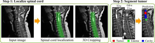
Spinal cord tumors lead to neurological morbidity and mortality. Being able to obtain morphometric quantification (size, location, growth rate) of the tumor, edema, and cavity can result in improved monitoring and treatment planning. Such quantification requires the segmentation of these structures into three separate classes. However, manual segmentation of three-dimensional structures is time consuming, tedious and prone to intra- and inter-rater variability, motivating the development of automated methods. Here, we tailor a model adapted to the spinal cord tumor segmentation task. Data were obtained from 343 patients using gadolinium-enhanced T1-weighted and T2-weighted MRI scans with cervical, thoracic, and/or lumbar coverage. The dataset includes the three most common intramedullary spinal cord tumor types: astrocytomas, ependymomas, and hemangioblastomas. The proposed approach is a cascaded architecture with U-Net-based models that segments tumors in a two-stage process: locate and label. The model first finds the spinal cord and generates bounding box coordinates. The images are cropped according to this output, leading to a reduced field of view, which mitigates class imbalance. The tumor is then segmented. The segmentation of the tumor, cavity, and edema (as a single class) reached 76.7 ± 1.5% of Dice score and the segmentation of tumors alone reached 61.8 ± 4.0% Dice score. The true positive detection rate was above 87% for tumor, edema, and cavity. To the best of our knowledge, this is the first fully automatic deep learning model for spinal cord tumor segmentation. The multiclass segmentation pipeline is available in the Spinal Cord Toolbox (https://spinalcordtoolbox.com/). It can be run with custom data on a regular computer within seconds.
中文翻译:
基于深度学习的 MRI 自动多类髓内脊髓肿瘤分割
脊髓肿瘤导致神经系统的发病率和死亡率。能够获得肿瘤、水肿和空腔的形态计量量化(大小、位置、生长速率)可以改善监测和治疗计划。这种量化需要将这些结构分成三个独立的类别。然而,三维结构的手动分割既费时又乏味,而且容易出现评分者内和评分者间的变异性,这推动了自动化方法的发展。在这里,我们定制了一个适合脊髓肿瘤分割任务的模型。数据来自 343 名患者,使用钆增强 T1 加权和 T2 加权 MRI 扫描覆盖颈椎、胸椎和/或腰椎。该数据集包括三种最常见的髓内脊髓肿瘤类型:星形细胞瘤、室管膜瘤、和血管母细胞瘤。所提出的方法是具有基于 U-Net 的模型的级联架构,该模型在两个阶段的过程中分割肿瘤:定位和标记。该模型首先找到脊髓并生成边界框坐标。根据此输出裁剪图像,导致视野减小,从而减轻类不平衡。然后分割肿瘤。肿瘤、空腔和水肿(作为单一类别)的分割达到了 Dice 评分的 76.7±1.5%,单独的肿瘤分割达到了 Dice 评分的 61.8±4.0%。肿瘤、水肿、空洞的真阳性检出率均在87%以上。据我们所知,这是第一个用于脊髓肿瘤分割的全自动深度学习模型。脊髓工具箱中提供了多类分割管道 ( 所提出的方法是具有基于 U-Net 的模型的级联架构,该模型在两个阶段的过程中分割肿瘤:定位和标记。该模型首先找到脊髓并生成边界框坐标。根据此输出裁剪图像,导致视野减小,从而减轻类不平衡。然后分割肿瘤。肿瘤、空腔和水肿(作为单一类别)的分割达到了 Dice 评分的 76.7±1.5%,单独的肿瘤分割达到了 Dice 评分的 61.8±4.0%。肿瘤、水肿、空洞的真阳性检出率均在87%以上。据我们所知,这是第一个用于脊髓肿瘤分割的全自动深度学习模型。脊髓工具箱中提供了多类分割管道 ( 所提出的方法是具有基于 U-Net 的模型的级联架构,该模型在两个阶段的过程中分割肿瘤:定位和标记。该模型首先找到脊髓并生成边界框坐标。根据此输出裁剪图像,导致视野减小,从而减轻类不平衡。然后分割肿瘤。肿瘤、空腔和水肿(作为单一类别)的分割达到了 Dice 评分的 76.7±1.5%,单独的肿瘤分割达到了 Dice 评分的 61.8±4.0%。肿瘤、水肿、空洞的真阳性检出率均在87%以上。据我们所知,这是第一个用于脊髓肿瘤分割的全自动深度学习模型。脊髓工具箱中提供了多类分割管道 (https://spinalcordtoolbox.com/)。它可以在几秒钟内在普通计算机上使用自定义数据运行。


 京公网安备 11010802027423号