当前位置:
X-MOL 学术
›
NMR Biomed.
›
论文详情
Our official English website, www.x-mol.net, welcomes your
feedback! (Note: you will need to create a separate account there.)
Volumetric coronary endothelial function assessment: a feasibility study exploiting stack-of-stars 3D cine MRI and image-based respiratory self-gating
NMR in Biomedicine ( IF 2.7 ) Pub Date : 2021-07-21 , DOI: 10.1002/nbm.4589 Gabriele Bonanno 1, 2 , Robert G Weiss 1, 2 , Davide Piccini 3 , Jérôme Yerly 4, 5 , Sahar Soleimani 2 , Li Pan 6 , Xiaoming Bi 7 , Allison G Hays 1 , Matthias Stuber 4, 5 , Michael Schär 2
NMR in Biomedicine ( IF 2.7 ) Pub Date : 2021-07-21 , DOI: 10.1002/nbm.4589 Gabriele Bonanno 1, 2 , Robert G Weiss 1, 2 , Davide Piccini 3 , Jérôme Yerly 4, 5 , Sahar Soleimani 2 , Li Pan 6 , Xiaoming Bi 7 , Allison G Hays 1 , Matthias Stuber 4, 5 , Michael Schär 2
Affiliation
|
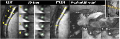
Abnormal coronary endothelial function (CEF), manifesting as depressed vasoreactive responses to endothelial-specific stressors, occurs early in atherosclerosis, independently predicts cardiovascular events, and responds to cardioprotective interventions. CEF is spatially heterogeneous along a coronary artery in patients with atherosclerosis, and thus recently developed and tested non-invasive 2D MRI techniques to measure CEF may not capture the extent of changes in CEF in a given coronary artery. The purpose of this study was to develop and test the first volumetric coronary 3D MRI cine method for assessing CEF along the proximal and mid-coronary arteries with isotropic spatial resolution and in free-breathing. This approach, called 3D-Stars, combines a 6 min continuous, untriggered golden-angle stack-of-stars acquisition with a novel image-based respiratory self-gating method and cardiac and respiratory motion-resolved reconstruction. The proposed respiratory self-gating method agreed well with respiratory bellows and center-of-k-space methods. In healthy subjects, 3D-Stars vessel sharpness was non-significantly different from that by conventional 2D radial in proximal segments, albeit lower in mid-portions. Importantly, 3D-Stars detected normal vasodilatation of the right coronary artery in response to endothelial-dependent isometric handgrip stress in healthy subjects. Coronary artery cross-sectional areas measured using 3D-Stars were similar to those from 2D radial MRI when similar thresholding was used. In conclusion, 3D-Stars offers good image quality and shows feasibility for non-invasively studying vasoreactivity-related lumen area changes along the proximal coronary artery in 3D during free-breathing.
中文翻译:

体积冠状动脉内皮功能评估:利用星栈 3D 电影 MRI 和基于图像的呼吸自门控的可行性研究
冠状动脉内皮功能异常(CEF),表现为对内皮特异性应激源的血管反应性反应降低,发生在动脉粥样硬化早期,独立预测心血管事件,并对心脏保护干预措施做出反应。动脉粥样硬化患者的 CEF 沿冠状动脉具有空间异质性,因此最近开发和测试的用于测量 CEF 的非侵入性 2D MRI 技术可能无法捕获给定冠状动脉中 CEF 的变化程度。本研究的目的是开发和测试第一个体积冠状动脉 3D MRI 电影方法,用于以各向同性空间分辨率和自由呼吸的方式评估近端和中部冠状动脉的 CEF。这种称为 3D-Stars 的方法将 6 分钟连续、未触发的黄金角恒星堆栈采集与新颖的基于图像的呼吸自门控方法以及心脏和呼吸运动分辨重建相结合。所提出的呼吸自门控方法与呼吸风箱和k空间中心方法非常一致。在健康受试者中,3D-Stars 血管清晰度与近端节段的传统 2D 径向血管清晰度没有显着差异,尽管中间部分较低。重要的是,3D-Stars 检测到健康受试者右冠状动脉的正常血管舒张,以响应内皮依赖性等长握力压力。当使用相似的阈值时,使用 3D-Stars 测量的冠状动脉横截面积与 2D 径向 MRI 测量的冠状动脉横截面积相似。总之,3D-Stars 提供了良好的图像质量,并显示了在自由呼吸期间以 3D 方式非侵入性研究沿近端冠状动脉的血管反应性相关管腔面积变化的可行性。
更新日期:2021-07-21
中文翻译:
体积冠状动脉内皮功能评估:利用星栈 3D 电影 MRI 和基于图像的呼吸自门控的可行性研究
冠状动脉内皮功能异常(CEF),表现为对内皮特异性应激源的血管反应性反应降低,发生在动脉粥样硬化早期,独立预测心血管事件,并对心脏保护干预措施做出反应。动脉粥样硬化患者的 CEF 沿冠状动脉具有空间异质性,因此最近开发和测试的用于测量 CEF 的非侵入性 2D MRI 技术可能无法捕获给定冠状动脉中 CEF 的变化程度。本研究的目的是开发和测试第一个体积冠状动脉 3D MRI 电影方法,用于以各向同性空间分辨率和自由呼吸的方式评估近端和中部冠状动脉的 CEF。这种称为 3D-Stars 的方法将 6 分钟连续、未触发的黄金角恒星堆栈采集与新颖的基于图像的呼吸自门控方法以及心脏和呼吸运动分辨重建相结合。所提出的呼吸自门控方法与呼吸风箱和k空间中心方法非常一致。在健康受试者中,3D-Stars 血管清晰度与近端节段的传统 2D 径向血管清晰度没有显着差异,尽管中间部分较低。重要的是,3D-Stars 检测到健康受试者右冠状动脉的正常血管舒张,以响应内皮依赖性等长握力压力。当使用相似的阈值时,使用 3D-Stars 测量的冠状动脉横截面积与 2D 径向 MRI 测量的冠状动脉横截面积相似。总之,3D-Stars 提供了良好的图像质量,并显示了在自由呼吸期间以 3D 方式非侵入性研究沿近端冠状动脉的血管反应性相关管腔面积变化的可行性。


 京公网安备 11010802027423号