Computer Methods and Programs in Biomedicine ( IF 4.9 ) Pub Date : 2021-07-21 , DOI: 10.1016/j.cmpb.2021.106279 Gözde Dursun 1 , Saurabh Balkrishna Tandale 1 , Rutwik Gulakala 1 , Jörg Eschweiler 2 , Mersedeh Tohidnezhad 3 , Bernd Markert 1 , Marcus Stoffel 1
|
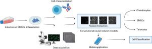
Background and objective: The use of automated systems for image recognition is highly preferred for regenerative medicine applications to evaluate stem cell differentiation early in the culturing state with non-invasive methodologies instead of invasive counterparts. Bone marrow-derived mesenchymal stem cells (BMSCs) are able to differentiate into desired cell phenotypes, and thereby promise a proper cell source for tendon regeneration. The therapeutic success of stem cell therapy requires cellular characterization prior to the implantation of cells. The foremost problem is that traditional characterization techniques require cellular material which would be more useful for cell therapy, complex laboratory procedures, and human expertise. Convolutional neural networks (CNNs), a class of deep neural networks, have recently made great improvements in image-based classifications, recognition, and detection tasks. We, therefore, aim to develop a potential CNN model in order to recognize differentiated stem cells by learning features directly from image data of unlabelled cells.
Methods: The differentiation of bone marrow mesenchymal stem cells (BMSCs) into tenocytes was induced with the treatment of bone morphogenetic protein-12 (BMP-12). Following the treatment and incubation step, the phase-contrast images of cells were obtained and immunofluorescence staining has been applied to characterize the differentiated state of BMSCs. CNN models were developed and trained with the phase-contrast cell images. The comparison of CNN models was performed with respect to prediction performance and training time. Moreover, we have evaluated the effect of image enhancement method, data augmentation, and fine-tuning training strategy to increase classification accuracy of CNN models. The best model was integrated into a mobile application.
Results: All the CNN models can fit the biological data extracted from immunofluorescence characterization. CNN models enable the cell classification with satisfactory accuracies. The best result in terms of accuracy and training time is achieved by the model proposed based on Inception-ResNet V2 trained from scratch using image enhancement and data augmentation strategies (96.80%, 434.55 sec).
Conclusion: Our study reveals that the CNN models show good performance by identifying stem cell differentiation. Importantly this technique provides a faster and real-time tool in comparison to traditional methods enabling the adjustment of culture conditions during cultivation to improve the yield of therapeutic stem cells.
中文翻译:
基于细胞形态识别肌腱分化的卷积神经网络的开发
背景和目标:在再生医学应用中,高度推荐使用自动化系统进行图像识别,以在培养状态的早期使用非侵入性方法而不是侵入性方法评估干细胞分化。骨髓间充质干细胞 (BMSCs) 能够分化成所需的细胞表型,从而有望为肌腱再生提供合适的细胞来源。干细胞疗法的治疗成功需要在细胞植入之前进行细胞表征。最重要的问题是,传统的表征技术需要细胞材料,这对细胞治疗、复杂的实验室程序和人类专业知识更有用。卷积神经网络(CNN),一类深度神经网络,最近在基于图像的分类、识别和检测任务方面取得了很大的进步。因此,我们的目标是开发一个潜在的 CNN 模型,以便通过直接从未标记细胞的图像数据中学习特征来识别分化的干细胞。
方法:通过骨形态发生蛋白12(BMP-12)诱导骨髓间充质干细胞(BMSCs)向肌腱细胞分化。在处理和孵育步骤之后,获得细胞的相差图像,并应用免疫荧光染色来表征 BMSCs 的分化状态。CNN 模型是使用相衬细胞图像开发和训练的。CNN 模型的比较是在预测性能和训练时间方面进行的。此外,我们评估了图像增强方法、数据增强和微调训练策略的效果,以提高 CNN 模型的分类精度。最佳模型已集成到移动应用程序中。
结果:所有CNN模型都可以拟合从免疫荧光表征中提取的生物数据。CNN 模型使细胞分类具有令人满意的准确性。在准确性和训练时间方面的最佳结果是基于 Inception-ResNet V2 提出的模型,使用图像增强和数据增强策略从头开始训练(96.80%,434.55 秒)。
结论:我们的研究表明,CNN 模型通过识别干细胞分化显示出良好的性能。重要的是,与传统方法相比,该技术提供了一种更快、更实时的工具,可以在培养过程中调整培养条件,以提高治疗性干细胞的产量。


 京公网安备 11010802027423号