当前位置:
X-MOL 学术
›
Environ. Sci.: Nano
›
论文详情
Our official English website, www.x-mol.net, welcomes your
feedback! (Note: you will need to create a separate account there.)
Understanding the toxicity mechanism of CuO nanoparticles: the intracellular view of exposed earthworm cells
Environmental Science: Nano ( IF 5.8 ) Pub Date : 2021-07-21 , DOI: 10.1039/d1en00080b Natividad Isabel Navarro Pacheco 1, 2, 3, 4, 5 , Radka Roubalova 1, 2, 3 , Jiri Dvorak 1, 2, 3 , Oldrich Benada 1, 2, 3 , Dominik Pinkas 3, 6, 7, 8, 9 , Olga Kofronova 1, 2, 3 , Jaroslav Semerad 1, 2, 3, 10, 11 , Martin Pivokonsky 3, 12, 13 , Tomas Cajthaml 1, 2, 3, 10, 11 , Martin Bilej 1, 2, 3 , Petra Prochazkova 1, 2, 3
Environmental Science: Nano ( IF 5.8 ) Pub Date : 2021-07-21 , DOI: 10.1039/d1en00080b Natividad Isabel Navarro Pacheco 1, 2, 3, 4, 5 , Radka Roubalova 1, 2, 3 , Jiri Dvorak 1, 2, 3 , Oldrich Benada 1, 2, 3 , Dominik Pinkas 3, 6, 7, 8, 9 , Olga Kofronova 1, 2, 3 , Jaroslav Semerad 1, 2, 3, 10, 11 , Martin Pivokonsky 3, 12, 13 , Tomas Cajthaml 1, 2, 3, 10, 11 , Martin Bilej 1, 2, 3 , Petra Prochazkova 1, 2, 3
Affiliation
|
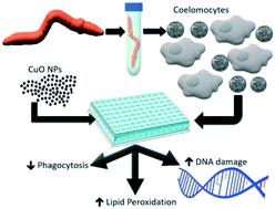
Copper oxide nanoparticles (CuO NPs) are widely used in industry. Once released, they can enter the soil system and endanger organisms living in this environment. Therefore, monitoring the NP impact on soil organisms and identification of suitable biomarkers associated with NP pollution are required. In this study, immune effector cells of the earthworm Eisenia andrei, amoebocytes, were exposed to environmentally relevant sublethal concentrations of CuO NPs (1, 10, and 100 μg mL−1 of Cu) and their impact on the cellular and subcellular levels, as well as on the mRNA levels of molecules involved in the defense reactions, was assessed in vitro. CuO NPs decreased the viability of both amoebocyte subpopulations by 40% at the highest concentration tested (100 μg mL−1 of Cu). Further, CuO NPs caused significant attenuation of the phagocytic function of hyaline amoebocytes after 6 and 24 hours of exposure, by 37 and 25%, respectively. The concentration of the lipid peroxidation subproduct, malondialdehyde, was 10 times elevated in cells exposed to CuO NPs (100 μg mL−1 of Cu) after 6 hours of exposure. We hypothesize that malondialdehyde may induce DNA breaks, cell cycle arrest, and subsequent cell death. Electron microscopy showed the interaction between CuO NPs and immune effector cells, amoebocytes. Moreover, aggregates of CuO NPs were shown to be engulfed and located in the cytoplasm of these cells. However, data from all experiments indicate that the observed effects of CuO NPs on earthworm coelomocytes were caused mainly by the dissolved Cu2+ ions derived from nanoparticles (NPs). The determination of effective parameters such as oxidative stress, immune reactivity, and genotoxicity would provide valuable comprehension and data for environmental assessment of NP impact on soil organisms.
中文翻译:

了解CuO纳米粒子的毒性机制:暴露的蚯蚓细胞的细胞内视图
氧化铜纳米粒子(CuO NPs)在工业中被广泛使用。一旦释放,它们就会进入土壤系统并危及生活在该环境中的生物。因此,需要监测 NP 对土壤生物的影响并确定与 NP 污染相关的合适生物标志物。在这项研究中,蚯蚓Eisenia andrei 的免疫效应细胞(变形虫细胞)暴露于环境相关亚致死浓度的 CuO NPs(1、10 和 100 μg mL -1的 Cu)及其对细胞和亚细胞水平的影响,如以及参与防御反应的分子的 mRNA 水平,在体外进行了评估. 在测试的最高浓度(100 μg mL -1 Cu)下,CuO NPs 将两种变形细胞亚群的活力降低了 40% 。此外,在暴露 6 小时和 24 小时后,CuO NPs 导致透明变形细胞的吞噬功能显着减弱,分别为 37% 和 25%。在暴露于 CuO NPs (100 μg mL -1Cu) 暴露 6 小时后。我们假设丙二醛可能导致 DNA 断裂、细胞周期停滞和随后的细胞死亡。电子显微镜显示 CuO NPs 与免疫效应细胞、变形细胞之间的相互作用。此外,CuO NPs 的聚集体被证明被吞没并位于这些细胞的细胞质中。然而,所有实验的数据表明,观察到的 CuO NPs 对蚯蚓体腔细胞的影响主要是由来自纳米颗粒 (NPs)的溶解的 Cu 2+离子引起的。氧化应激、免疫反应性和遗传毒性等有效参数的确定将为 NP 对土壤生物影响的环境评估提供有价值的理解和数据。
更新日期:2021-07-21
中文翻译:
了解CuO纳米粒子的毒性机制:暴露的蚯蚓细胞的细胞内视图
氧化铜纳米粒子(CuO NPs)在工业中被广泛使用。一旦释放,它们就会进入土壤系统并危及生活在该环境中的生物。因此,需要监测 NP 对土壤生物的影响并确定与 NP 污染相关的合适生物标志物。在这项研究中,蚯蚓Eisenia andrei 的免疫效应细胞(变形虫细胞)暴露于环境相关亚致死浓度的 CuO NPs(1、10 和 100 μg mL -1的 Cu)及其对细胞和亚细胞水平的影响,如以及参与防御反应的分子的 mRNA 水平,在体外进行了评估. 在测试的最高浓度(100 μg mL -1 Cu)下,CuO NPs 将两种变形细胞亚群的活力降低了 40% 。此外,在暴露 6 小时和 24 小时后,CuO NPs 导致透明变形细胞的吞噬功能显着减弱,分别为 37% 和 25%。在暴露于 CuO NPs (100 μg mL -1Cu) 暴露 6 小时后。我们假设丙二醛可能导致 DNA 断裂、细胞周期停滞和随后的细胞死亡。电子显微镜显示 CuO NPs 与免疫效应细胞、变形细胞之间的相互作用。此外,CuO NPs 的聚集体被证明被吞没并位于这些细胞的细胞质中。然而,所有实验的数据表明,观察到的 CuO NPs 对蚯蚓体腔细胞的影响主要是由来自纳米颗粒 (NPs)的溶解的 Cu 2+离子引起的。氧化应激、免疫反应性和遗传毒性等有效参数的确定将为 NP 对土壤生物影响的环境评估提供有价值的理解和数据。


 京公网安备 11010802027423号