Brain Research ( IF 2.7 ) Pub Date : 2021-07-18 , DOI: 10.1016/j.brainres.2021.147585 Logan J Voss 1 , Maxence Plouviez 2 , Nicola Whittle 1
|
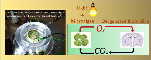
Hypoxic brain injury is a leading cause of loss of quality of life globally for which there are currently no effective treatments. There has been increasing interest in incorporating photosynthesising agents into hypoxic tissue as a mechanism for in situ oxygen delivery, independent of vascular perfusion. To date this has not been tested in the brain.
The oxygen production capacity of Chlamydomonas reinhardtii microalgal cultures was measured in artificial cerebrospinal fluid (aCSF) in benchtop assays and in cortical slices in situ. Cortical slice function was quantified by measuring the length, frequency and amplitude of seizure-like event (SLE) activity — in conventionally oxygenated aCSF, C. reinhardtii cultures, unoxygenated and deoxygenated aCSF. The possibility of direct toxic algal effects was investigated by exposing slices to cultures for 5 h. An oxygen level of 25 mg.L−1 was achieved with C. reinhardtii in no-Mg aCSF. Slice SLE function was preserved in C. reinhardtii, without the need for supplemental oxygen. In contrast, functional parameters deteriorated in unoxygenated and deoxygenated aCSF. In the former, there was a 66% reduction in SLE frequency and a 37% reduction in event amplitude. In the latter, SLE activity ceased completely. No toxic algae effects were seen in slices exposed to cultures for 5 h.
These results confirm that C. reinhardtii oxygenation of aCSF can sustain cortical network activity — without tissue toxicity for the normal lifespan of an acute cortical slice. This study shows promise for the concept of photosynthesis as a mechanism for providing oxygen to rescue ischaemic avascularised brain tissue.
中文翻译:
基于微藻的光合策略为无血管小鼠脑组织供氧——一项体外概念验证研究
缺氧性脑损伤是全球生活质量下降的主要原因,目前尚无有效的治疗方法。人们越来越关注将光合作用剂结合到缺氧组织中作为原位氧气输送的机制,而与血管灌注无关。迄今为止,这还没有在大脑中进行过测试。
莱茵衣藻微藻培养物的产氧能力在人工脑脊液 (aCSF) 中的台式测定和原位皮质切片中进行了测量。通过测量癫痫样事件 (SLE) 活动的长度、频率和幅度来量化皮质切片功能——在常规含氧 aCSF、莱茵衣藻培养、未含氧和脱氧 aCSF 中。通过将切片暴露于培养物 5 小时来研究直接有毒藻类效应的可能性。在无镁 aCSF中用莱茵衣藻实现了25 mg.L -1的氧气水平。切片 SLE 功能保留在莱茵衣藻中,无需补充氧气。相比之下,功能参数在无氧和脱氧 aCSF 中恶化。在前者中,SLE 频率降低了 66%,事件幅度降低了 37%。在后者中,SLE 活动完全停止。在暴露于培养物 5 小时的切片中未观察到有毒藻类效应。
这些结果证实, aCSF 的莱茵衣藻氧合可以维持皮质网络活动——在急性皮质切片的正常寿命期间没有组织毒性。这项研究表明,光合作用的概念有望成为一种提供氧气来拯救缺血性无血管脑组织的机制。


 京公网安备 11010802027423号