当前位置:
X-MOL 学术
›
Phys. Rev. Appl.
›
论文详情
Our official English website, www.x-mol.net, welcomes your
feedback! (Note: you will need to create a separate account there.)
Conformational changes of a membrane protein determined by infrared difference spectroscopy beyond the diffraction limit
Physical Review Applied ( IF 3.8 ) Pub Date : 2021-07-20 , DOI: 10.1103/physrevapplied.16.014048 Raffaella Polito 1 , Maria Eleonora Temperini 1, 2 , Eglof Ritter 3, 4 , Ljiljana Puskar 3 , Ulrich Schade 3 , Matthias Broser 4 , Peter Hegemann 4 , Leonetta Baldassarre 1 , Michele Ortolani 1 , Valeria Giliberti 2
Physical Review Applied ( IF 3.8 ) Pub Date : 2021-07-20 , DOI: 10.1103/physrevapplied.16.014048 Raffaella Polito 1 , Maria Eleonora Temperini 1, 2 , Eglof Ritter 3, 4 , Ljiljana Puskar 3 , Ulrich Schade 3 , Matthias Broser 4 , Peter Hegemann 4 , Leonetta Baldassarre 1 , Michele Ortolani 1 , Valeria Giliberti 2
Affiliation
|
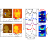
Optogenetics is a revolutionary method for studying neural activity by exploiting the optical stimulation of neurons made possible by artificial genetic expression of light-sensitive transmembrane “gate” proteins. Evidence of functional conformational changes of the gate proteins can be obtained by difference infrared (IR) absorption spectroscopy triggered by light stimuli. Here we investigate the effect on the photocycle of the prototype optogenetic protein channelrhodopsin (ChR2) of gold surfaces placed in close proximity to the protein molecules. In order to do this, we bring difference IR spectroscopy to the nanoscale, using a platform based on the coupling of a tunable mid-IR quantum cascade laser and an atomic force microscope (AFM). Sensitivity to individual subwavelength-sized membrane patches, embedding fewer than a hundred ChR2 molecules, is achieved by taking advantage of a plasmonic field enhancement in the nanogap between the gold-coated AFM tip and an ultraflat gold surface used as a sample support. We obtain relative difference absorption variations smaller than and benchmark nanospectroscopy difference spectra against those taken with the Fourier transform IR (FTIR) spectroscopic technique on the same sample. We identify distinct simultaneous processes in the ChR2 photocycle by performing singular value decomposition of the FTIR difference spectra, and use this procedure to compare IR nanospectroscopy data on individual membrane patches in contact with gold surfaces with those obtained on thick stacks of membrane patches unexposed to metal surfaces. ChR2 proteins maintain their gate function when placed in a 14-nm-wide gap between two gold surfaces, apart from minor modifications to their kinetics. Our results are relevant to optogenetic applications that require physical contact between nanoscale metallic probes and electrodes and the membrane of single neuronal cells. More generally, our work paves the way towards the spectroscopic study of transmembrane proteins at the nanoscale.
中文翻译:

红外差分光谱法测定的膜蛋白构象变化超出衍射极限
光遗传学是一种革命性的研究神经活动的方法,它利用光敏跨膜“门”蛋白的人工基因表达使神经元的光刺激成为可能。门蛋白功能构象变化的证据可以通过光刺激触发的差红外 (IR) 吸收光谱获得。在这里,我们研究了靠近蛋白质分子放置的金表面的原型光遗传蛋白通道视紫红质 (ChR2) 对光循环的影响。为了做到这一点,我们使用基于可调谐中红外量子级联激光器和原子力显微镜 (AFM) 耦合的平台,将差分红外光谱引入纳米级。对单个亚波长大小的膜贴片敏感,嵌入少于一百个 ChR2 分子,通过利用镀金 AFM 尖端和用作样品支撑的超平坦金表面之间的纳米间隙中的等离子体场增强来实现。我们获得的相对差异吸收变化小于和基准纳米光谱差异光谱与傅立叶变换红外 (FTIR) 光谱技术对同一样品的光谱进行对比。我们通过对 FTIR 差异光谱进行奇异值分解来识别 ChR2 光循环中不同的同时过程,并使用此程序将与金表面接触的单个膜贴片的 IR 纳米光谱数据与未暴露于金属的厚膜贴片堆叠上获得的数据进行比较表面。ChR2 蛋白在放置在两个金表面之间 14 纳米宽的间隙中时保持其门功能,除了对其动力学进行微小修改。我们的结果与需要纳米级金属探针和电极与单个神经元细胞膜之间的物理接触的光遗传学应用相关。更普遍,
更新日期:2021-07-20
中文翻译:
红外差分光谱法测定的膜蛋白构象变化超出衍射极限
光遗传学是一种革命性的研究神经活动的方法,它利用光敏跨膜“门”蛋白的人工基因表达使神经元的光刺激成为可能。门蛋白功能构象变化的证据可以通过光刺激触发的差红外 (IR) 吸收光谱获得。在这里,我们研究了靠近蛋白质分子放置的金表面的原型光遗传蛋白通道视紫红质 (ChR2) 对光循环的影响。为了做到这一点,我们使用基于可调谐中红外量子级联激光器和原子力显微镜 (AFM) 耦合的平台,将差分红外光谱引入纳米级。对单个亚波长大小的膜贴片敏感,嵌入少于一百个 ChR2 分子,通过利用镀金 AFM 尖端和用作样品支撑的超平坦金表面之间的纳米间隙中的等离子体场增强来实现。我们获得的相对差异吸收变化小于和基准纳米光谱差异光谱与傅立叶变换红外 (FTIR) 光谱技术对同一样品的光谱进行对比。我们通过对 FTIR 差异光谱进行奇异值分解来识别 ChR2 光循环中不同的同时过程,并使用此程序将与金表面接触的单个膜贴片的 IR 纳米光谱数据与未暴露于金属的厚膜贴片堆叠上获得的数据进行比较表面。ChR2 蛋白在放置在两个金表面之间 14 纳米宽的间隙中时保持其门功能,除了对其动力学进行微小修改。我们的结果与需要纳米级金属探针和电极与单个神经元细胞膜之间的物理接触的光遗传学应用相关。更普遍,


 京公网安备 11010802027423号