当前位置:
X-MOL 学术
›
Microsc. Res. Tech.
›
论文详情
Our official English website, www.x-mol.net, welcomes your
feedback! (Note: you will need to create a separate account there.)
Scanning electron microscopy of the palatine mucosa with connective tissue papillae of the Egyptian red fox (Vulpes vulpes aegyptiaca, Linnaeus, 1758)
Microscopy Research and Technique ( IF 2.0 ) Pub Date : 2021-07-20 , DOI: 10.1002/jemt.23877 Mohamed A A Mahdy 1 , Elsayed S I Mohammed 2
Microscopy Research and Technique ( IF 2.0 ) Pub Date : 2021-07-20 , DOI: 10.1002/jemt.23877 Mohamed A A Mahdy 1 , Elsayed S I Mohammed 2
Affiliation
|
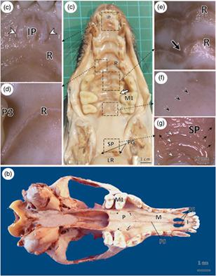
The present study investigated the morphological, as well as the histological features of the Egyptian red fox's palate, in addition to the three-dimensional characteristics of the connective tissue papillae (CTP) of the palate by scanning electron microscopy (SEM). The hard palate was narrow rostrally and its width increased caudally. The maximum width was located at the caudal border of the upper fourth premolar tooth. The incisive papilla was represented by a rounded median elevation surrounded on each side by a groove on which the oral openings of the incisive ducts opened. The rostral part of the hard palate had 9–10 caudally concave transverse palatine ridges while the caudal part appeared smooth without ridges. The palatine raphe was indistinct rostrally but formed a groove caudally. By SEM, the palatine ridges had low projections. Different microplicae systems were present on the epithelial surface of the incisive papilla, palatine rugae, interrugal areas, and the smooth part. The CTP of the incisive papilla, palatine ridges, and soft palate were conical-shaped, cylindrical-shaped, and parallel serrated ridges, respectively. Histologically, the hard palate was lined by a cornified stratified squamous epithelium resting on a dense connective layer of lamina propria while the soft palate was lined by a noncornified stratified squamous epithelium. The palatine salivary glands were present in the smooth part of the hard palate and the soft palate. The information presented in the current study might serve as a reference guide for the interpretation of pathological conditions of the palate of red fox.
中文翻译:

埃及红狐(Vulpes vulpes aegyptiaca,Linnaeus,1758)的带有结缔组织乳头的腭粘膜的扫描电子显微镜
本研究通过扫描电子显微镜 (SEM) 研究了埃及红狐上颚的形态学和组织学特征,以及上颚结缔组织乳头 (CTP) 的三维特征。硬腭口吻狭窄,尾端宽度增加。最大宽度位于上第四前磨牙的尾缘。切牙乳头由圆形的正中隆起表示,每侧被凹槽包围,切牙导管的口腔开口在凹槽上打开。硬腭的喙部有9-10个尾侧凹的横向腭脊,而尾部则显得光滑无脊。腭中缝在嘴上不明显,但在尾部形成一个凹槽。通过 SEM,腭脊具有低突起。切牙乳头、腭皱、皱间区和光滑部分的上皮表面存在不同的微皱襞系统。切牙乳头、腭脊和软腭的CTP分别为圆锥形、圆柱形和平行锯齿状脊。组织学上,硬腭由位于固有层致密结缔层上的角化复层鳞状上皮排列,而软腭由非角化复层鳞状上皮排列。腭唾液腺存在于硬腭和软腭的光滑部分。当前研究中提供的信息可作为解释赤狐上颚病理状况的参考指南。
更新日期:2021-07-20
中文翻译:
埃及红狐(Vulpes vulpes aegyptiaca,Linnaeus,1758)的带有结缔组织乳头的腭粘膜的扫描电子显微镜
本研究通过扫描电子显微镜 (SEM) 研究了埃及红狐上颚的形态学和组织学特征,以及上颚结缔组织乳头 (CTP) 的三维特征。硬腭口吻狭窄,尾端宽度增加。最大宽度位于上第四前磨牙的尾缘。切牙乳头由圆形的正中隆起表示,每侧被凹槽包围,切牙导管的口腔开口在凹槽上打开。硬腭的喙部有9-10个尾侧凹的横向腭脊,而尾部则显得光滑无脊。腭中缝在嘴上不明显,但在尾部形成一个凹槽。通过 SEM,腭脊具有低突起。切牙乳头、腭皱、皱间区和光滑部分的上皮表面存在不同的微皱襞系统。切牙乳头、腭脊和软腭的CTP分别为圆锥形、圆柱形和平行锯齿状脊。组织学上,硬腭由位于固有层致密结缔层上的角化复层鳞状上皮排列,而软腭由非角化复层鳞状上皮排列。腭唾液腺存在于硬腭和软腭的光滑部分。当前研究中提供的信息可作为解释赤狐上颚病理状况的参考指南。


 京公网安备 11010802027423号